File:X-ray of healthy lungs taken at the Institute of Tropical Medicine in Antwerp, on 9 December 2016 (1).tif

Original file (11,555 × 9,279 pixels, file size: 86.8 MB, MIME type: image/tiff)
Captions
Captions
Summary[edit]
| Warning | The original file is very high-resolution. It might not load properly or could cause your browser to freeze when opened at full size. |
|---|
| Description |
English: X-ray of healthy lungs taken at the Institute of Tropical Medicine in Antwerp, on 9 December 2016 (1).
Text visible on the radiograph: USER ID:Jverbeken@Agfa Healthcare ♦ INSTITUUT VOOR TROPISCHE GENEESKUNDE - ANTWERPEN ♦ VERHEYEN, VINCENT MIA, ♦ 1992.12.22 ♦ 2016.12.09 ♦ 11:41:46 ♦ PRINT DATE: 20161209 TIME: 11:48:07 Medical analysis by Dr. J. Verbeken (combined judgement, also based on another radiograph of the subject's lungs): The following exams were executed: ♦ Chest X-ray: ♦ The diaphragm is well delineated and the costo-phrenic sinuses are free. ♦ Broncho-vascular hilar shadows. ♦ Normal cardiac shadow. ♦ Free apices. ♦ Normal aeration of both lungs. ♦ No arguments for alveolar infiltrations. ♦ Normal aspe[c]t of the thorax skeleton. ♦ Conclusion: ♦ No infiltrations. No pleural effusions. No masses. ♦ No tuberculosis. ♦ No other contageous lung diseases. Administration number: 07004479; Reference: 161209005. |
| Date | |
| Source | Scan of original X-ray. |
| Author | N.a. Radiology by Dr. Johan Verbeken. Scan and image editing by Vincent Mia Edie Verheyen. |
| Camera location | | View this and other nearby images on: OpenStreetMap |
|---|
| This is a retouched picture, which means that it has been digitally altered from its original version. Modifications: Adjusted contrast and lightning levels. Modifications made by Vincent Mia Edie Verheyen.
|
Licensing[edit]
| This file is ineligible for copyright and therefore in the public domain, because it is a technical image created as part of a standard medical diagnostic procedure. No creative element rising above the threshold of originality was involved in its production. See Meta:Wikilegal/Copyright of Medical Imaging for details.
|
 |
In the Public Domain as this is not copyrightable content. For a discussion, see https://commons.wikimedia.org/wiki/Commons:Requests_for_comment/Xrays. The subject hereby revokes all privacy concerns.
File history
Click on a date/time to view the file as it appeared at that time.
| Date/Time | Thumbnail | Dimensions | User | Comment | |
|---|---|---|---|---|---|
| current | 13:50, 9 December 2016 |  | 11,555 × 9,279 (86.8 MB) | Vanished user lkjhgfdsazxcvbnm (talk | contribs) | Increased contrast, fiddled with lightning levels. |
| 13:34, 9 December 2016 | 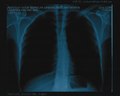 | 11,555 × 9,279 (306.78 MB) | Vanished user lkjhgfdsazxcvbnm (talk | contribs) | User created page with UploadWizard |
You cannot overwrite this file.
File usage on Commons
The following page uses this file: