Category:Medical X-rays (images)
Jump to navigation
Jump to search

English: Medical X-rays
Français : Radiographies médicales
| NO WIKIDATA ID FOUND! Search for Medical X-rays (images) on Wikidata | |
| Upload media |
Subcategories
This category has the following 15 subcategories, out of 15 total.
+
-
- Images by Arthur Schuster (47 F)
A
C
I
N
O
- X-rays of osteoblastoma (1 F)
P
- X-rays with body piercings (4 F)
S
Media in category "Medical X-rays (images)"
The following 200 files are in this category, out of 230 total.
(previous page) (next page)-
 X- ray with ear ring.jpg 3,584 × 2,213; 3.35 MB
-

-
 111-SC-16187 - NARA - 55192476.jpg 8,405 × 5,669; 19.89 MB
-
 12881 2018 682 Fig1 HTML.png 685 × 825; 227 KB
-
 12920 2023 1472 Fig3 HTML.webp 685 × 841; 58 KB
-
 21518 lores.jpg 700 × 866; 36 KB
-
 3848035360 9a1402df7a bFractureCrâne.jpg 2,805 × 2,239; 491 KB
-
 738a35ed1b3843a28a8be69409b5f6 thumb.jpg 135 × 176; 5 KB
-
 8822753.fig.001.svg 348 × 348; 1.29 MB
-
 A woman's narrow pelvis with child's head stuck in the middl Wellcome V0014960.jpg 2,479 × 3,094; 2.34 MB
-
 A woman's pelvis after a pubiotomy - to widen the birth cana Wellcome V0014957.jpg 2,224 × 3,138; 2.15 MB
-
 The A B C of the X rays (IA abcofxrays00mead 0).pdf 789 × 1,150, 206 pages; 13.63 MB
-
 The ABC of the X-rays (IA abcxrays00meadgoog).pdf 843 × 1,168, 210 pages; 6.59 MB
-
 Afiliado, (sharpening) o filtrado pasa alta.png 1,184 × 508; 266 KB
-
 Ahlbaeck.jpg 658 × 694; 253 KB
-
 American quarterly of roentgenology (1906) (14777160173).jpg 2,300 × 3,596; 3.13 MB
-
 ANAEURISMA0003.jpg 512 × 816; 42 KB
-
 Anatomischer Anzeiger (1886) (18174086465).jpg 1,746 × 2,668; 1.09 MB
-
 Ankylosing spondylitis of lumbosacral spine.png 958 × 702; 581 KB
-
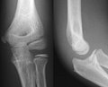 Ap lateral elbow.jpg 660 × 532; 30 KB
-
 AP radiograph demonstrating companion shadow of the clavicle.jpg 2,016 × 2,106; 261 KB
-
 AP Whole spine with piercing.jpg 2,418 × 5,738; 3.61 MB
-

-
 Artificial Intelligence for X-ray inspection.png 576 × 672; 332 KB
-

-

-
 Barium swallow of malignancy oesophagus 01.jpg 935 × 675; 33 KB
-
 Barium swallow of malignancy oesophagus 02.JPG 935 × 675; 38 KB
-
 Barium swallow of malignancy oesophagus 03.JPG 935 × 675; 36 KB
-
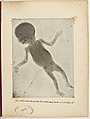 Beş aylık cenin, 1896.jpg 1,509 × 2,000; 453 KB
-
 BotonAortico.jpg 669 × 567; 148 KB
-
 Bound feet (X-ray).jpg 3,709 × 2,892; 4.11 MB
-
 Boy chest x-ray pic.jpg 2,305 × 3,096; 1.24 MB
-
 Boy pelvic x-ray pic.jpg 2,221 × 3,082; 1.54 MB
-
 Caput-sourcil angle.jpg 1,143 × 704; 106 KB
-
 Cardinal vowels-Jones x-ray.jpg 588 × 577; 118 KB
-
 Cayton.jpg 643 × 656; 119 KB
-
 Cervical x ray.jpg 1,080 × 1,280; 51 KB
-
 Chest X-ray of a normal lung with old left 5th and 6th ribs fractures.jpg 4,608 × 3,456; 3.03 MB
-

-
 Chest X-ray PA erect showing left upper lobe cavitation.png 3,456 × 4,608; 15.47 MB
-
 Chest x-ray plain film normal.jpg 1,753 × 1,885; 335 KB
-
 Chest X-ray showing left lower zone lung mass behind the heart.jpg 842 × 1,024; 342 KB
-
 Chest X-ray showing neurofibromatosis with bilateral pneumonia.jpg 790 × 702; 80 KB
-
 Chest X-ray showing presence of free air under left diaphragm.png 1,792 × 1,024; 2.28 MB
-
 Chest X-Ray.jpg 657 × 776; 114 KB
-
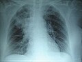
 Chest X-ray.jpg 4,113 × 4,613; 7.58 MB
-
 Chest XR of HAPE.png 996 × 724; 1.23 MB
-
 Deacon McGuire hand x-ray.jpg 336 × 376; 41 KB
-
 Die Röntgentechnik - Lehrbuch für Ärzte und Studierende (1906) (14571727577).jpg 1,566 × 1,568; 486 KB
-
 Die Röntgentechnik - Lehrbuch für Ärzte und Studierende (1906) (14755846824).jpg 1,936 × 1,674; 397 KB
-
 Disc 44.jpg 1,185 × 457; 56 KB
-
 Discograph degenerative disc disease L5-S1.jpg 1,488 × 1,128; 260 KB
-
 Dislocated Finger XRay.png 390 × 441; 31 KB
-
 Dogs paw y-ray ap.jpg 269 × 773; 24 KB
-

-
 Esostosi gamba.jpg 2,532 × 3,378; 1.47 MB
-

-
 Fractured Fibula.jpg 3,024 × 4,032; 5.37 MB
-
 Frauenhand MET DP262472.jpg 1,886 × 3,874; 1.55 MB
-
 Fuss eines 17 jährigen Jünglings MET DP262475.jpg 2,847 × 3,889; 1.88 MB
-
 Gerendertes DVT mit Nervdarstellung klein.jpg 250 × 179; 16 KB
-
 Gerendertes DVT mit Nervdarstellung.jpg 500 × 358; 19 KB
-
 Haarverlaengerungen im Roentgenbild - 001.jpg 1,025 × 1,116; 126 KB
-
 Hand eines 4 jährigen Kindes MET DP262474.jpg 1,904 × 3,884; 1.48 MB
-
 Hand eines 8 jährigen Mädchens MET DP262473.jpg 2,235 × 3,884; 1.77 MB
-
 Hand X-ray one.jpg 3,504 × 2,336; 655 KB
-
 Hand X-ray two.jpg 3,504 × 2,336; 1.05 MB
-
 Gary Simmons' CT scan.jpg 830 × 1,174; 172 KB
-
 Hueso trigono II.jpg 403 × 403; 14 KB
-
 Hypochondropl 17J W.jpeg 439 × 999; 175 KB
-
 Información de la composición de la imagen.png 884 × 672; 625 KB
-
 Interpretation of dental and maxillary roentgenograms (1918) (14571372020).jpg 1,268 × 1,250; 372 KB
-
 Interpretation of dental and maxillary roentgenograms (1918) (14571564328).jpg 1,272 × 1,246; 205 KB
-
 Interpretation of dental and maxillary roentgenograms (1918) (14754894291).jpg 1,284 × 1,268; 166 KB
-
 Interstate medical journal (1917) (14597078289).jpg 1,580 × 2,552; 367 KB
-
 Interstate medical journal (1917) (14597100499).jpg 1,408 × 1,116; 99 KB
-
 Interstate medical journal (1917) (14597118678).jpg 1,414 × 1,042; 152 KB
-
 Interstate medical journal (1917) (14597121738).jpg 1,380 × 1,054; 112 KB
-
 Interstate medical journal (1917) (14597250157).jpg 1,274 × 1,056; 131 KB
-
 Interstate medical journal (1917) (14760770846).jpg 1,394 × 1,052; 125 KB
-
 Interstate medical journal (1917) (14760771296).jpg 1,430 × 2,204; 369 KB
-
 Interstate medical journal (1917) (14780599001).jpg 1,610 × 2,682; 353 KB
-
 Interstate medical journal (1917) (14781387534).jpg 1,584 × 2,716; 372 KB
-
 Interstate medical journal (1917) (14783676105).jpg 1,250 × 1,584; 196 KB
-
 Interstate medical journal (1917) (14803620633).jpg 1,732 × 2,478; 317 KB
-
 Journal of radiology (1921) (14571476138).jpg 2,358 × 2,884; 392 KB
-
 Knee plain X-ray.jpg 1,936 × 2,595; 2.15 MB
-
 Küntschers Femurnagel von 1940 (1987).JPG 556 × 1,232; 244 KB
-
 The bones of the wrist of Mrs Herries; two views. Wellcome L0047930.jpg 4,616 × 3,815; 2.6 MB
-
 Lateral Chest X-Ray.jpg 704 × 819; 92 KB
-

-
 Lunatummalazie AP.jpg 269 × 222; 35 KB
-
 Lung X-ray.jpg 2,293 × 2,293; 2.42 MB
-
 LWS ap.jpg 521 × 924; 92 KB
-
 MBq digital-radiograph.jpg 1,207 × 403; 65 KB
-
 Medizinische Panoramaaufnahme.jpg 1,574 × 902; 435 KB
-
 Mesurements Hindfoot Alignment View.jpg 184 × 375; 34 KB
-
 Mummy image.jpg 1,615 × 5,045; 1.06 MB
-
 My Back.jpg 958 × 645; 114 KB
-
 Neumonia.jpg 245 × 184; 6 KB
-
 Normal chest radiograph which is under-inspired and is rotated.jpg 4,608 × 3,456; 4.28 MB
-
 Normal neonatal chest x-ray.jpg 714 × 644; 65 KB
-

-

-
 OtherForComparison.jpg 579 × 490; 42 KB
-

-
 PAVLOVIC MARIJA CR 20130724 90028 1.jpg 2,328 × 2,928; 323 KB
-
 Pelvis of Albert Fish (X-ray).jpg 217 × 208; 14 KB
-
 Pelvis of E. Welch, showing disease of right hip and disloca Wellcome L0026321.jpg 1,616 × 1,212; 617 KB
-
 Plain radiograph of the right hip .jpg 330 × 599; 21 KB
-
 PMC3202142 CRIM2011-527569.001.png 512 × 415; 166 KB
-
 Pnemoni-görüntüleme.jpg 586 × 336; 107 KB
-
 Pointmaker Annotation of Digital XRay.jpg 1,500 × 1,355; 622 KB
-
 Principles of modern biology (1964) (20744080881).jpg 1,228 × 1,410; 524 KB
-
 ProtuberanceEruption 19190529 MeudonSpectroheliograph.jpg 1,952 × 1,665; 639 KB
-
 Provincial Reconstruction Team, Forward Surgical Team Docs DVIDS246359.jpg 2,700 × 1,793; 2.71 MB
-
 PSM V48 D857 Photo of early x ray.jpg 1,860 × 2,735; 1.05 MB
-
 PSM V50 D678 Effect of 12 second fluorescent screen exposure.jpg 2,798 × 1,729; 801 KB
-
 PSM V56 D0681 Dental x ray of a ten year old patient.png 1,225 × 1,270; 301 KB
-
 PSM V56 D0682 X ray photo of an elbow joint.png 1,626 × 666; 233 KB
-
 Radio jambe plus courte.jpg 2,448 × 3,264; 2.05 MB
-
 Radiografia-dental.jpg 3,264 × 2,448; 384 KB
-
 Radiografía de Luis Zegers - 22 marzo 1896.jpg 949 × 1,407; 821 KB
-
 Radiografía dental panorámica.jpg 775 × 452; 111 KB
-
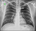
 Radiograph showing IVC Filter Fracture.jpg 320 × 161; 9 KB
-
 Radiography of hand 40 days after hand transplant.jpg 676 × 1,333; 100 KB
-
 Radiography of the right and left arm after hand transplant.jpg 729 × 833; 76 KB
-
 Right Foot of Male human 38 years old side view.png 1,366 × 2,278; 1.47 MB
-
 Right Foot of Male human 38 years old top view.png 1,266 × 2,210; 906 KB
-

-
-

-

-
-

-

-

-
-
-
-
 Rx de tórax F-Cabello.jpg 875 × 788; 483 KB
-
 RX on femur after curettage surgery 2.jpg 2,417 × 2,428; 735 KB
-
 RX on femur after curettage surgery.jpg 2,192 × 2,301; 563 KB
-
 Saltzmann-Aufnahme, junger Erwachsener.jpg 856 × 1,484; 276 KB
-
 Schlittenprothese.jpg 862 × 592; 37 KB
-
 Slide 1.png 1,080 × 1,500; 554 KB
-
 Small Lumbar Scoliosis Curve with Pelvic Torque.png 712 × 830; 396 KB
-
 STAR$20Welser.jpg 263 × 350; 11 KB
-
 Stress-view-of-ankle-with-deltoid-ligament-tear.jpg 1,129 × 1,535; 141 KB
-
 Su caso.jpg 2,106 × 758; 179 KB
-
 SVG of x-ray of a hand in 1896.svg 2,514 × 2,090; 607 KB
-
 The American annual of photography (1922) (14594948168).jpg 1,926 × 2,396; 1.38 MB
-

-
 The left and right knee-joints of Frank Burgess, probably a Wellcome L0026319.jpg 1,198 × 1,542; 671 KB
-

-

-

-

-

-

-

-

-

-

-

-

-

-
 The Röntgen rays in medical work (1899) (14570308648).jpg 2,094 × 3,372; 1.85 MB
-
 The Röntgen rays in medical work (1899) (14733888186).jpg 1,704 × 3,544; 482 KB
-
 The Röntgen rays in medical work (1899) (14733934726).jpg 2,194 × 3,658; 2.34 MB
-
 The Röntgen rays in medical work (1899) (14776809683).jpg 2,106 × 3,672; 1.92 MB
-
 The Röntgen rays in medical work (1907) (14571065087).jpg 2,800 × 1,634; 849 KB
-
 The San Pedro Mountain Mummy.jpg 505 × 287; 43 KB
-
 Transformaciones de intensidad CT.png 802 × 452; 250 KB
-

-
 Unbehandelter Perthes.jpg 990 × 2,386; 764 KB
-
 Vegetationsstörungen und Systemerkrankungen der Knochen (1899) (14755801154).jpg 1,696 × 2,902; 782 KB
-
 Vessel-X container.JPG 2,796 × 2,230; 925 KB
-
 World War I radiography amputee.jpg 1,024 × 751; 183 KB
-
 WPV X-Ray1.jpg 1,506 × 1,224; 187 KB
-
 X ray internal fixation leg fracture.jpg 2,448 × 3,057; 3.3 MB
-
 X ray of Fore arm ap and lat view.jpg 1,060 × 1,109; 92 KB
-
 X-ray dogs paw ml.jpg 457 × 412; 16 KB
-
 X-ray dosage in treatment and radiography (1922) (14758066605).jpg 1,388 × 1,114; 554 KB
-
 X-ray General Illustration.jpg 1,792 × 3,117; 1.18 MB
-

-
 X-ray observations for foreign bodies and their localisation (1920) (14757764862).jpg 2,912 × 1,674; 619 KB
-
 X-ray of a knee joint, showing shrapnel Wellcome V0030078.jpg 3,504 × 2,044; 2.25 MB
-

-
 X-ray of a rib-cage, showing shrapnel Wellcome V0030080.jpg 2,988 × 2,351; 1.94 MB
-
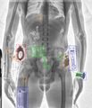
 X-ray of abdomen with perforated IUD.jpg 1,876 × 2,247; 863 KB
-
 X-ray of an arm, showing a broken bone. Wellcome V0030074.jpg 3,344 × 2,096; 2.11 MB
-
 X-Ray of foot joint.jpeg 3,264 × 2,448; 1.16 MB
-
 X-ray of hand deformed by osteo-arthritis. Wellcome M0019124.jpg 2,835 × 3,892; 2.57 MB
-

-
-
 X-ray of Idiopathic interstital pneumonia.jpg 600 × 495; 22 KB
-
 X-ray of slate pneumoconiosis sufferer's lungs. Wellcome L0029726.jpg 1,248 × 1,625; 456 KB
-
 X-ray of Trinket Swallowed by a Child (FDA010) (6946959167).jpg 2,048 × 1,604; 195 KB


_o_filtrado_pasa_alta.png)

_(14777160173).jpg)

_(18174086465).jpg)


_(14570647579).jpg)
_(14570694618).jpg)


.jpg)


,_National_Museum_of_Health_and_Medicine.jpg)


_(14571727577).jpg)
_(14755846824).jpg)


_(14571372020).jpg)
_(14571564328).jpg)
_(14754894291).jpg)
_(14597078289).jpg)
_(14597100499).jpg)
_(14597118678).jpg)
_(14597121738).jpg)
_(14597250157).jpg)
_(14760770846).jpg)
_(14760771296).jpg)
_(14780599001).jpg)
_(14781387534).jpg)
_(14783676105).jpg)
_(14803620633).jpg)
_(14571476138).jpg)

.JPG)


,_National_Museum_of_Health_and_Medicine_(253519649).jpg)


_(14570916187).jpg)

.jpg)


_(20744080881).jpg)


_(14571440600).jpg)
_(14571455180).jpg)
_(14571504849).jpg)
_(14571575159).jpg)
_(14571682737).jpg)
_(14571761337).jpg)
_(14754984221).jpg)
_(14757846782).jpg)
_(14777998643).jpg)
_(14778080133).jpg)
_(14778125713).jpg)


_(14594948168).jpg)
.jpg)

_(14570846270).jpg)
_(14570848690).jpg)
_(14570856149).jpg)
_(14571084797).jpg)
_(14734594846).jpg)
_(14754411311).jpg)
_(14754450311).jpg)
_(14755241344).jpg)
_(14757235232).jpg)
_(14757506905).jpg)
_(14757552605).jpg)
_(14777445863).jpg)
_(14758028672).jpg)
_(14570308648).jpg)
_(14733888186).jpg)
_(14733934726).jpg)
_(14776809683).jpg)
_(14571065087).jpg)

-_2014-05-27_04-55.jpg)
_(14755801154).jpg)


_(14758066605).jpg)


_(14757764862).jpg)


_(6946959167).jpg)
&filefrom=X-Ray+on+Left+Hand+Post-Proximal+Row+Carpectomy+1.jpg#mw-category-media)
_(14571371407).jpg)
_(14757821865).jpg)