File:Lax 33014 elife-33014-fig2-v1.jpg
Jump to navigation
Jump to search

Size of this preview: 798 × 600 pixels. Other resolutions: 320 × 240 pixels | 639 × 480 pixels | 1,022 × 768 pixels | 1,280 × 962 pixels | 1,500 × 1,127 pixels.
Original file (1,500 × 1,127 pixels, file size: 534 KB, MIME type: image/jpeg)
Captions
Captions
Add a one-line explanation of what this file represents
Summary
[edit]| Description |
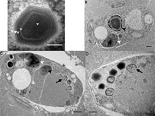
English: Ultrastructure of Bodo saltans virus (BsV, Theiavirus salishense) particles and replication.
(A) Mature BsV virion: DNA containing core is surrounded by two putative membranous layers. The capsid consists of at least two proteinaceous layers. The bright halo hints to the presence of short (~40 nm) fibers as observed in Acantha polyphaga memivirus (ApMV). The top vertex of the virion contains a possible stargate structure. (Scale bar = 100 nm) (B) Healthy Bodo saltans cell: Nucleus with nucleolus and heterochromatin structures (Back arrow head) and kinetoplast genome (white arrow head) are clearly visible. (Scale bar = 500 nm) (C) Cell of B. saltans 24 hr post-BsV infection: Most subcellular compartments of healthy cells have been displaced by the virus factory now taking up a third of the cell. Virion production is directed toward the periphery of the cell (black arrow). Kinetoplast genome remains intact (white arrow head) while the nuclear genome is degraded (black arrow head; Scale bar = 500 nm) (D) BsV virion assembly and maturation: Lipid vesicles migrate through the virion factory where capsid proteins attach for the proteinaceous shell. Vesicles burst and accumulate at the virus factory periphery where the capsid assembly completes (black arrow). Once the capsid is assembled, the virion is filled with the genome and detaches from the virus factory. Internal structures develop inside the virion in the cell’s periphery where mature virions accumulate until the host cell bursts (Scale bar = 500 nm).
Deutsch: (A) zeigt ein reifes Virion von Bodo saltans virus (BsV). (B) zeigt eine gesunde Zelle von Bodo saltans. (C) und (D) zeigen den Zustand nach der Infektion, die Assemblierung und die Reifung der Viruspartikel in einer Zelle von B. saltans. |
| Date | |
| Source |
Fig. 2 at https://elifesciences.org/articles/33014/figures at https://elifesciences.org/articles/33014 The kinetoplastid-infecting Bodo saltans virus (BsV), a window into the most abundant giant viruses in the sea. In: eLife 2018;7:e33014 doi:10.7554/eLife.33014 |
| Author | Christoph M. Deeg, Cheryl-Emiliane T. Chow, Curtis A. Suttle |
| Other versions |
    |


Licensing
[edit]
This file is licensed under the Creative Commons Attribution-Share Alike 4.0 International license.
- You are free:
- to share – to copy, distribute and transmit the work
- to remix – to adapt the work
- Under the following conditions:
- attribution – You must give appropriate credit, provide a link to the license, and indicate if changes were made. You may do so in any reasonable manner, but not in any way that suggests the licensor endorses you or your use.
- share alike – If you remix, transform, or build upon the material, you must distribute your contributions under the same or compatible license as the original.
File history
Click on a date/time to view the file as it appeared at that time.
| Date/Time | Thumbnail | Dimensions | User | Comment | |
|---|---|---|---|---|---|
| current | 08:44, 14 April 2021 | | 1,500 × 1,127 (534 KB) | Ernsts (talk | contribs) | Uploaded a work by Christoph M. Deeg, Cheryl-Emiliane T. Chow, Curtis A. Suttle from https://elifesciences.org/articles/33014/figures at https://elifesciences.org/articles/33014 The kinetoplastid-infecting Bodo saltans virus (BsV), a window into the most abundant giant viruses in the sea. In: eLife 2018;7:e33014 doi:10.7554/eLife.33014 50px|class=noviewer with UploadWizard |
You cannot overwrite this file.
File usage on Commons
The following 9 pages use this file:
- File:BsV from eLife.jpg
- File:Lax 33014 elife-33014-fig2-figsupp1-v1.jpg
- File:Lax 33014 elife-33014-fig2A-v1.jpg
- File:Lax 33014 elife-33014-fig2B-v1.jpg
- File:Lax 33014 elife-33014-fig2C-v1.jpg
- File:Lax 33014 elife-33014-fig2D-v1.jpg
- File:Lax 33014 elife-33014-fig3-v1.jpg
- File:Lax 33014 elife-33014-fig6C-v1.jpg
- File:Lax 33014 elife-33014-fig7-v1.jpg
File usage on other wikis
The following other wikis use this file:
- Usage on de.wikipedia.org
- Usage on en.wikipedia.org
- Usage on species.wikimedia.org