File:F29-01-9780123846846-Rudiviridae-Fig1-SRV.png
Jump to navigation
Jump to search

Size of this preview: 800 × 441 pixels. Other resolutions: 320 × 177 pixels | 640 × 353 pixels | 1,024 × 565 pixels | 1,550 × 855 pixels.
Original file (1,550 × 855 pixels, file size: 516 KB, MIME type: image/png)
Captions
Captions
Add a one-line explanation of what this file represents
Summary
[edit]
| Description |
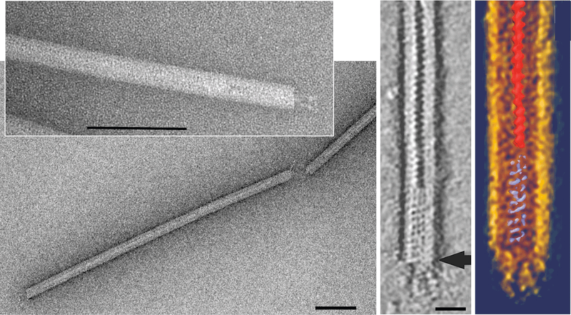
English: Rudiviridae. (Left panel) Negative contrast electron micrographs of virions of Stygiolobus rod-shaped virus. The bars correspond to 200 nm. (Right panels) Electron tomography image of the virion: horizontal slice (0.7) showing the accumulated stain in the central part, and visualization of the data using Amira software. With an arrow is indicated a point of attachment of the three tail fibers. The scale bar represents 50 nm. |
| Date | |
| Source | https://talk.ictvonline.org/cfs-file/__key/communityserver-wikis-components-files/00-00-00-00-21/f29_2D00_01_2D00_9780123846846.png at https://talk.ictvonline.org/ictv-reports/ictv_9th_report/dsdna-viruses-2011/w/dsdna_viruses/132/rudiviridae-figures ICTV 9th Report (2011) Rudiviridae - Figures: Fig. 1 |
| Author | International Committee on Taxonomy of Viruses (ICTV): Modified from Vestergaard et al. (2008). J. Bacteriol., 190, 6837-6845. |
| Other versions |
  |
Licensing
[edit]
This file is licensed under the Creative Commons Attribution-Share Alike 4.0 International license.
- You are free:
- to share – to copy, distribute and transmit the work
- to remix – to adapt the work
- Under the following conditions:
- attribution – You must give appropriate credit, provide a link to the license, and indicate if changes were made. You may do so in any reasonable manner, but not in any way that suggests the licensor endorses you or your use.
- share alike – If you remix, transform, or build upon the material, you must distribute your contributions under the same or compatible license as the original.
File history
Click on a date/time to view the file as it appeared at that time.
| Date/Time | Thumbnail | Dimensions | User | Comment | |
|---|---|---|---|---|---|
| current | 10:51, 24 March 2021 | | 1,550 × 855 (516 KB) | Ernsts (talk | contribs) | Uploaded a work by International Committee on Taxonomy of Viruses (ICTV): Modified from Vestergaard ''et al.'' (2008). J. Bacteriol., 190, 6837-6845. from https://talk.ictvonline.org/cfs-file/__key/communityserver-wikis-components-files/00-00-00-00-21/f29_2D00_01_2D00_9780123846846.png at https://talk.ictvonline.org/ictv-reports/ictv_9th_report/dsdna-viruses-2011/w/dsdna_viruses/132/rudiviridae-figures ICTV 9th Report (2011) Rudiviridae - Figures: Fig. 1 with UploadWizard |
You cannot overwrite this file.
File usage on Commons
The following 2 pages use this file:
File usage on other wikis
The following other wikis use this file:
- Usage on de.wikipedia.org