File:F12-01-9780123846846.png
Jump to navigation
Jump to search

Size of this preview: 600 × 600 pixels. Other resolutions: 240 × 240 pixels | 480 × 480 pixels | 768 × 768 pixels | 1,024 × 1,024 pixels | 2,047 × 2,047 pixels.
Original file (2,047 × 2,047 pixels, file size: 2.43 MB, MIME type: image/png)
Captions
Captions
Add a one-line explanation of what this file represents
Summary
[edit]| Description |
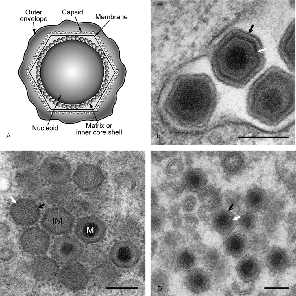
English: (A) Diagram of extracellular ASFV virions showing nucleoid, matrix or inner core shell, capsid and lipid envelopes. (B) EM image of extracellular virions. Black arrow is outer envelope, white arrow is virus membrane. Bar=200 nm. The preparation method was standard chemical fixation for EM. (C) EM image of intracellular virions. IM=immature virion, M=mature virion. Black arrow is capsid protein, white arrow is virus membrane. Bar=200 nm. Preparation method was high pressure freezing followed by freeze substitution. (D) EM image of intracellular virions. Black arrow is capsid protein, white arrow is virus membrane. Bar=200 nm. Preparation method was thawed cryo-sections stained with uranyl acetate. |
| Date | |
| Source | Fig. 1 at https://talk.ictvonline.org/ictv-reports/ictv_9th_report/dsdna-viruses-2011/w/dsdna_viruses/100/asfarviridae-figures dsDNA Viruses > Family: Afarviridae. ICTV 9th Report (2011) |
| Author | Dixon, L.K., Alonso, C., Escribano, J.M., Martins, C., Revilla, Y., Salas, M.L. and Takamatsu, H.. Original by Pippa Hawes |
| Other versions |

Licensing
[edit]
This file is licensed under the Creative Commons Attribution-Share Alike 4.0 International license.
- You are free:
- to share – to copy, distribute and transmit the work
- to remix – to adapt the work
- Under the following conditions:
- attribution – You must give appropriate credit, provide a link to the license, and indicate if changes were made. You may do so in any reasonable manner, but not in any way that suggests the licensor endorses you or your use.
- share alike – If you remix, transform, or build upon the material, you must distribute your contributions under the same or compatible license as the original.
File history
Click on a date/time to view the file as it appeared at that time.
| Date/Time | Thumbnail | Dimensions | User | Comment | |
|---|---|---|---|---|---|
| current | 19:22, 20 April 2021 | | 2,047 × 2,047 (2.43 MB) | Ernsts (talk | contribs) | Uploaded a work by Dixon, L.K., Alonso, C., Escribano, J.M., Martins, C., Revilla, Y., Salas, M.L. and Takamatsu, H.. Original by Pippa Hawes from Fig. 1 at https://talk.ictvonline.org/ictv-reports/ictv_9th_report/dsdna-viruses-2011/w/dsdna_viruses/100/asfarviridae-figures dsDNA Viruses > Family: ''Afarviridae''. ICTV 9th Report (2011) with UploadWizard |
You cannot overwrite this file.
File usage on Commons
The following page uses this file:
File usage on other wikis
The following other wikis use this file:
- Usage on de.wikipedia.org