File:41598 2018 31638 Fig9 HTML.webp
Jump to navigation
Jump to search

Size of this PNG preview of this WEBP file: 784 × 600 pixels. Other resolutions: 314 × 240 pixels | 628 × 480 pixels | 1,004 × 768 pixels | 1,280 × 979 pixels | 1,650 × 1,262 pixels.
Original file (1,650 × 1,262 pixels, file size: 82 KB, MIME type: image/webp)
Captions
Captions
Add a one-line explanation of what this file represents
Summary
[edit]
| Description |
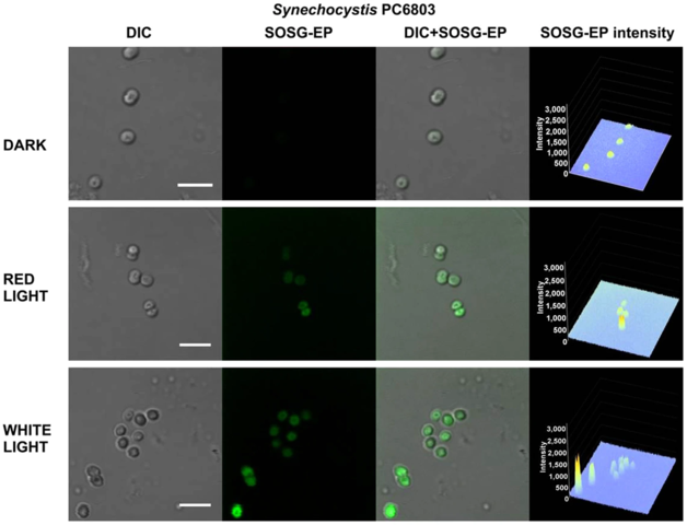
English: Effect of red and white light on singlet oxygen imaging in Synechocystis sp. PCC 6803 cells. Synechocystis cells were treated in 50 μM SOSG for 30 min at 37 °C either in dark or exposed to red/white light. For each treatment following images are presented (from left to right): Nomarski DIC, SOSG-EP fluorescence (λem = 505–525 nm), combined channel and integral distribution of the signal intensity within the sample (Z-axis represents the levels of brightness for each pixel, ranging between 0 and 3200). Bar represents 5 µm. |
| Date | |
| Source | Fig. 9 at https://www.nature.com/articles/s41598-018-31638-5 Singlet oxygen imaging using fluorescent probe Singlet Oxygen Sensor Green in photosynthetic organisms. In: nature Scientific Reports volume 8, Article number: 13685 doi:10.1038/s41598-018-31638-5 |
| Author | Ankush Prasad, Michaela Sedlářová, Pavel Pospíšil |
| Other versions |
  |
Licensing
[edit]
This file is licensed under the Creative Commons Attribution-Share Alike 4.0 International license.
- You are free:
- to share – to copy, distribute and transmit the work
- to remix – to adapt the work
- Under the following conditions:
- attribution – You must give appropriate credit, provide a link to the license, and indicate if changes were made. You may do so in any reasonable manner, but not in any way that suggests the licensor endorses you or your use.
- share alike – If you remix, transform, or build upon the material, you must distribute your contributions under the same or compatible license as the original.
File history
Click on a date/time to view the file as it appeared at that time.
| Date/Time | Thumbnail | Dimensions | User | Comment | |
|---|---|---|---|---|---|
| current | 16:36, 14 May 2021 | | 1,650 × 1,262 (82 KB) | Ernsts (talk | contribs) | Uploaded a work by Ankush Prasad, Michaela Sedlářová, Pavel Pospíšil from Fig. 9 at https://www.nature.com/articles/s41598-018-31638-5 Singlet oxygen imaging using fluorescent probe Singlet Oxygen Sensor Green in photosynthetic organisms. In: nature Scientific Reports volume 8, Article number: 13685 doi:10.1038/s41598-018-31638-5 with UploadWizard |
You cannot overwrite this file.
File usage on Commons
The following 2 pages use this file: