Category:Mikael Häggström/Micrographs of the gastrointestinal tract
Jump to navigation
Jump to search

These images were created by Mikael Häggström, M.D.
- User info
- Reusing images
Media in category "Mikael Häggström/Micrographs of the gastrointestinal tract"
The following 112 files are in this category, out of 112 total.
-
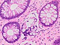
 Acute suppurative appendicitis with perforation.jpg 2,941 × 3,105; 2.79 MB
-
 Anorectal mucosa with hemorrhoid.jpg 2,048 × 1,532; 1.09 MB
-
 Beta-catenin immunohistochemistry in gastrointestinal stromal tumor (GIST).jpg 1,205 × 905; 257 KB
-
 Colorectal adenocarcinoma, not otherwise specified.jpg 1,705 × 1,781; 1.87 MB
-
 Counting Ki-67 index in immunohistochemistry.jpg 1,546 × 700; 295 KB
-
 Counting Ki-67 index in immunohistochemistry.svg 447 × 202; 261 KB
-
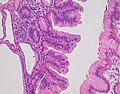
 Cross-section histology of small intestinal villi of the terminal ileum.jpg 2,048 × 1,532; 1.4 MB
-
 Dark, light, mantle and marginal zones of a secondary follicle, original.jpg 2,048 × 1,532; 709 KB
-
 Dark, light, mantle and marginal zones of a secondary follicle.png 1,313 × 979; 1.88 MB
-
 Duodenal biopsy with chronic active peptic duodenitis.jpg 2,048 × 1,532; 733 KB
-
 Enterobius vermicularis - intermediate magnification.jpg 2,048 × 1,532; 329 KB
-
 Enterobius vermicularis - low magnification.jpg 1,161 × 1,361; 323 KB
-
 Enterobius vermicularis egg.jpg 603 × 417; 58 KB
-
 Foveolar cells - crop.jpg 463 × 181; 20 KB
-
 Foveolar cells.jpg 2,080 × 1,536; 666 KB
-
 Gross pathology of metastatic melanoma to small intestine.jpg 1,541 × 1,881; 895 KB
-
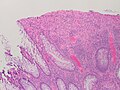
 Histology of a normal colonic mucosal epithelium.jpg 1,493 × 1,199; 460 KB
-
 Histology of an eosinophil in esophageal epithelium.jpg 547 × 487; 60 KB
-
 Histology of paneth cells, annotated.jpg 1,257 × 1,117; 367 KB
-
 Histology of paneth cells, original.jpg 2,048 × 1,532; 474 KB
-
 Histology of subepithelial lymphoid aggregate of the colon.jpg 2,730 × 2,042; 1.49 MB
-
 Histopathology of a crypt abscess.jpg 857 × 781; 234 KB
-
 Histopathology of a degenerated crypt of chronic inactive colitis.jpg 2,048 × 1,532; 464 KB
-
 Histopathology of a fundic glad polyp, low magnification.jpg 1,821 × 1,441; 592 KB
-
 Histopathology of a fundic gland polyp with dysplasia.jpg 1,891 × 1,479; 734 KB
-
 Histopathology of a gastric hyperplastic polyp, high magnification.jpg 2,080 × 1,536; 832 KB
-
 Histopathology of a mucosal fold.jpg 2,048 × 1,532; 415 KB
-
 Histopathology of a pedunculated colonic lipoma.jpg 2,281 × 3,753; 1.7 MB
-
 Histopathology of a well-differentiated neuroendocrine tumor on a stomach biopsy.jpg 1,461 × 1,329; 726 KB
-

-
 Histopathology of acute and chronic inflammation of the gastro-esophageal junction.jpg 1,536 × 2,080; 728 KB
-
 Histopathology of acute intraluminal inflammation of the appendix.jpg 2,048 × 1,532; 662 KB
-
 Histopathology of appendicitis.jpg 441 × 1,050; 201 KB
-

-
 Histopathology of Barrett's esophagus, annotated.jpg 2,047 × 1,799; 828 KB
-
 Histopathology of Barrett's esophagus, original.jpg 2,048 × 1,800; 711 KB
-
 Histopathology of cholelithiasis.jpg 1,417 × 1,069; 232 KB
-
 Histopathology of chronic appendicitis - low magnification.jpg 2,897 × 2,841; 2.35 MB
-
 Histopathology of chronic cholecystitis with gastric metaplasia.jpg 1,594 × 1,499; 730 KB
-
 Histopathology of colonic crypt branching.jpg 1,532 × 2,048; 802 KB
-
 Histopathology of colonic ulceration.jpg 2,048 × 1,532; 519 KB
-
 Histopathology of colorectal adenocarcinoma with lymphatic invasion.jpg 1,657 × 1,277; 640 KB
-
 Histopathology of crypt branching of colon.jpg 1,117 × 1,498; 459 KB
-
 Histopathology of degenerated crypt of chronic inactive colitis.jpg 2,048 × 1,532; 464 KB
-
 Histopathology of enterobius vermicularis eggs, HE stain.jpg 787 × 827; 125 KB
-
 Histopathology of eosinophilic esophagitis.jpg 2,048 × 1,532; 423 KB
-
 Histopathology of foveolar metaplasia of peptic duodenitis.jpg 1,661 × 1,197; 429 KB
-
 Histopathology of fundic gland polyp, high magnification, annotated.jpg 2,080 × 1,536; 495 KB
-
 Histopathology of fundic gland polyp, high magnification.jpg 2,080 × 1,536; 445 KB
-
 Histopathology of gastric tubular adenoma, high magnification.jpg 2,048 × 1,532; 519 KB
-
 Histopathology of gastric tubular adenoma, low magnification.jpg 1,792 × 1,193; 692 KB
-
 Histopathology of gland destruction in active colitis.jpg 1,531 × 1,161; 491 KB
-

-
 Histopathology of goblet cells and foveolar cells in incomplete Barrett's esophagus.jpg 2,048 × 1,532; 665 KB
-
 Histopathology of granuloma of colonic mucosa.jpg 1,465 × 1,313; 657 KB
-
 Histopathology of helicobacter pylori (annotated).jpg 259 × 223; 26 KB
-
 Histopathology of helicobacter pylori.jpg 259 × 278; 29 KB
-
 Histopathology of hemorrhoid.jpg 2,080 × 1,536; 865 KB
-
 Histopathology of high-grade dysplasia in a tubulovillous adenoma.jpg 2,048 × 1,532; 540 KB
-
 Histopathology of high-grade dysplasia in tubulovillous adenoma, annotated.png 2,048 × 1,532; 6.6 MB
-
 Histopathology of Histoplasma capsulatum, GMS stain.jpg 1,285 × 1,031; 208 KB
-
 Histopathology of histoplasma, HE stain.png 1,327 × 943; 1.54 MB
-
 Histopathology of hyperplastic polyp, tangential section.jpg 2,048 × 1,532; 699 KB
-
 Histopathology of hyperplastic polyp.jpg 2,080 × 1,536; 809 KB
-
 Histopathology of intestinal spirochetosis.jpg 1,817 × 1,847; 970 KB
-
 Histopathology of intraepithelial lymphocytes of the duodenum, annotated.jpg 1,213 × 789; 290 KB
-
 Histopathology of intravascular neutrophils.jpg 747 × 703; 187 KB
-
 Histopathology of leiomyoma of the ileocecal valve.jpg 3,072 × 2,048; 1.57 MB
-

-

-
 Histopathology of lymphocytic colitis, annotated.jpg 2,048 × 1,532; 713 KB
-
 Histopathology of lymphocytic colitis, high magnification.png 2,048 × 1,532; 3.69 MB
-
 Histopathology of lymphocytic colitis, low magnification.png 2,048 × 1,532; 4 MB
-
 Histopathology of lymphocytic colitis.jpg 2,048 × 1,532; 485 KB
-
 Histopathology of mild active gastritis, annotated.jpg 2,048 × 1,532; 588 KB
-
 Histopathology of mild active gastritis.jpg 2,048 × 1,532; 387 KB
-
 Histopathology of moderately differentiated esophageal adenocarcinoma.jpg 2,048 × 1,532; 509 KB
-
 Histopathology of mucosal plasma cell infiltrate in chronic gastritis.jpg 2,080 × 1,536; 810 KB
-
 Histopathology of neutrophilic cryptitis in active colitis, annotated.jpg 2,048 × 1,532; 540 KB
-
 Histopathology of neutrophilic cryptitis in active colitis, original.jpg 2,048 × 1,532; 450 KB
-
 Histopathology of patchy chronic peri-appendicitis.jpg 811 × 640; 188 KB
-
 Histopathology of peptic duodenitis - annotated.jpg 1,536 × 1,777; 651 KB
-
 Histopathology of peptic duodenitis.jpg 1,536 × 2,080; 719 KB
-
 Histopathology of periappendicitis.jpg 3,889 × 2,353; 1.99 MB
-
 Histopathology of reactive gastropathy, annotated.jpg 2,080 × 1,536; 741 KB
-
 Histopathology of reactive gastropathy.jpg 2,080 × 1,536; 735 KB
-
 Histopathology of reactive inflammatory changes in a hyperplastic polyp.jpg 1,917 × 1,061; 702 KB
-
 Histopathology of small crypt abscess.jpg 2,048 × 1,532; 457 KB
-
 Histopathology of submucosal lipoma.jpg 1,573 × 1,275; 658 KB
-
 Histopathology of traditional serrated adenoma, high magnification.jpg 2,080 × 1,536; 753 KB
-
 Histopathology of traditional serrated adenoma, low magnification.jpg 2,080 × 1,536; 946 KB
-
 Histopathology of ulcer in acute cholecystitis.jpg 2,397 × 2,325; 1.89 MB
-
 Histopathology Paneth cell metaplasia.jpg 2,048 × 1,532; 417 KB
-
 Histopathology venous congestion of the small intestine.jpg 2,080 × 1,536; 1.05 MB
-

-
 Lymph node metastasis from neuroendocrine tumor.jpg 1,159 × 969; 795 KB
-
 Melanoma resembling a squamous cell carcinoma.jpg 2,048 × 1,532; 820 KB
-
 Micrograph of colorectal carcinoma with dirty necrosis.jpg 773 × 659; 389 KB
-
 Micrograph of entry point of appendicular arteries.jpg 1,585 × 1,673; 1.14 MB
-
 Micrograph of mucosal disruption and mononuclear cells of the appendix.jpg 1,433 × 1,361; 901 KB
-
 Micrograph of tubulovillous adenoma with high grade dysplasia (annotated).jpg 1,861 × 2,105; 1.87 MB
-
 Micrograph of tubulovillous adenoma with high grade dysplasia.jpg 1,861 × 2,105; 1.88 MB
-
 Mitosis in a neuroendocrine tumor (original).jpg 2,048 × 1,532; 531 KB
-
 Mitosis in a neuroendocrine tumor.jpg 863 × 625; 151 KB
-
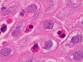
 Moderate superficial chronic gastritis.jpg 2,048 × 1,532; 1.21 MB
-
 Non-proliferative versus proliferative colonic crypts.jpg 2,080 × 1,536; 790 KB
-
 Normal versus dysplastic colonic crypt - low magnification.jpg 2,080 × 1,536; 943 KB
-
 Signet ring cell, original.jpg 1,029 × 855; 121 KB
-
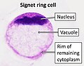 Signet ring cell.jpg 911 × 710; 115 KB
-
 Submucosal mononuclear cells in appendix - high magnification.jpg 2,080 × 1,536; 810 KB
-
 Synaptophysin immunohistochemistry of neuroendocrine tumor.jpg 1,665 × 1,793; 1.17 MB
-
 Trichuris trichiura eggs, including HE stain.jpg 401 × 265; 42 KB


.jpg)


_and_foveolar_cells_in_incomplete_Barrett%27s_esophagus.jpg)


.jpg)


.jpg)

.jpg)


