Category:Mikael Häggström/Micrographs
Jump to navigation
Jump to search

These images were created by Mikael Häggström, M.D.
- User info
- Reusing images
Subcategories
This category has the following 15 subcategories, out of 15 total.
M
- Mikael Häggström/Micrographs of the breast (1 P, 81 F)
Media in category "Mikael Häggström/Micrographs"
The following 161 files are in this category, out of 161 total.
-
 Adipose tissue with crumpling artifact due to insufficient fixation.jpg 2,048 × 1,532; 363 KB
-
 Apoptotic neutrophil with nuclear fragmentation.jpg 110 × 101; 11 KB
-
 Birefringence microscopy of gout, annotated.jpg 607 × 473; 62 KB
-
 Birefringence microscopy of gout, original.jpg 857 × 663; 91 KB
-
 Birefringence microscopy of pseudogout, annotated.jpg 1,745 × 1,581; 615 KB
-
 Birefringence microscopy of pseudogout.jpg 3,024 × 4,032; 1.5 MB
-
 Bone marrow cellularity in QuPath.png 1,421 × 597; 1.67 MB
-
 Calcium pyrophosphate dihydrate crystals without and with condenser, annotated.jpg 1,691 × 1,029; 437 KB
-
 Calcium pyrophosphate dihydrate crystals without and with condenser.jpg 1,693 × 1,033; 429 KB
-
 Calibration slide with rulers and grids.jpg 833 × 731; 151 KB
-
 Calibration slide.jpg 3,081 × 2,561; 2.06 MB
-
 CD117 (c-kit) stain of mixed malignant germ cell tumor - crop.png 482 × 374; 475 KB
-
 CD117 (c-kit) stain of mixed malignant germ cell tumor.png 1,073 × 905; 2.38 MB
-

-
 CK7 of metastatic carcinoma to lymph node.jpg 1,197 × 1,113; 234 KB
-
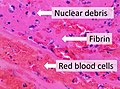 Composition of a fresh thrombus.jpg 2,041 × 1,521; 825 KB
-
 Cross-section of whipworm on microscopy, original.jpg 983 × 613; 151 KB
-
 Cross-section of whipworm on microscopy.jpg 849 × 773; 140 KB
-
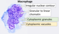 Cytology of a macrophage.png 1,097 × 629; 511 KB
-
 Cytology of normal mesothelium, original.jpg 2,048 × 1,532; 244 KB
-
 Cytology of normal mesothelium.jpg 2,269 × 2,109; 692 KB
-
 Cytology of reactive mesothelium, original.jpg 2,048 × 1,532; 305 KB
-
 Cytology of reactive mesothelium.jpg 2,125 × 1,581; 531 KB
-
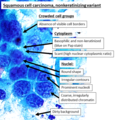 Cytopathology of nonkeratinizing squamous cell carcinoma.png 579 × 604; 432 KB
-
 Cytopathology of pleomorphic adenoma, original.jpg 2,048 × 1,532; 324 KB
-
 Cytopathology of pleomorphic adenoma.png 1,391 × 1,083; 2.02 MB
-
 Cytopathology of Warthin's tumor, original.jpg 1,528 × 2,050; 588 KB
-
 Cytopathology of Warthin's tumor.jpg 3,209 × 2,271; 953 KB
-
 Eosinophilic, basophilic, chromophobic and amphophilic staining.png 993 × 985; 1.05 MB
-
 Field of view diameter in microscopy.jpg 2,653 × 2,327; 1.1 MB
-
 Fine versus coarse chromatin.jpg 1,365 × 621; 181 KB
-
 Folding artifact on whole slide imaging of bone.png 1,103 × 913; 1.02 MB
-
 Gram negative rods and gram positive cocci 1.jpg 2,048 × 1,532; 244 KB
-
 Gram negative rods and gram positive cocci 2.jpg 2,048 × 1,532; 232 KB
-
 Gram positive coccus and gram negative rod.png 313 × 165; 46 KB
-
 Gram stain of a macrophage with ingested S epidermidis bacteria.jpg 2,048 × 1,532; 326 KB
-
 HE stained section of adenocarcinoma in peritoneal fluid.jpg 2,048 × 1,532; 342 KB
-
 Heterochromatic versus euchromatic nuclei.jpg 449 × 352; 60 KB
-
 High magnification histology of pars distalis of the anterior pituitary gland.png 2,080 × 1,536; 4.39 MB
-
 Histology of a lipoblast-like histiocyte in fat necrosis.jpg 2,048 × 1,532; 511 KB
-

-

-
 Histology of synovial membrane.jpg 2,048 × 1,237; 684 KB
-
 Histology of the mesothelial lining of a hernia sac.jpg 2,048 × 845; 313 KB
-
 Histology of vas deferens.jpg 2,048 × 1,532; 645 KB
-
 Histopatholgoy of acute gangrenous cholecystitis.jpg 2,048 × 1,532; 894 KB
-
 Histopathology of a bile duct hamartoma, high magnification.jpg 2,048 × 1,532; 476 KB
-
 Histopathology of a bile duct hamartoma, low magnification.jpg 1,561 × 1,393; 1.11 MB
-
 Histopathology of a biopsy of a melanoma metastasis to the brain.jpg 2,048 × 1,532; 470 KB
-
 Histopathology of a blood clot with postmortem bacterial growth.jpg 731 × 613; 150 KB
-
 Histopathology of a centroblast in follicular lymphoma.jpg 226 × 186; 27 KB
-
 Histopathology of a false diverticulum of the gallbladder.jpg 2,557 × 2,445; 1.52 MB
-
 Histopathology of a fibrolipoma.jpg 1,873 × 1,565; 1.44 MB
-
 Histopathology of a fresh thrombus, low magnification.jpg 2,048 × 1,532; 898 KB
-
 Histopathology of a fresh thrombus.jpg 2,048 × 1,532; 538 KB
-
 Histopathology of a hyperplastic polyp of the gallbladder.jpg 3,081 × 1,937; 1.43 MB
-
 Histopathology of a metastatic melanoma to a lymph node.jpg 2,048 × 1,532; 405 KB
-
 Histopathology of a reactive lymph node in cholecystitis.jpg 2,080 × 1,536; 1.34 MB
-
 Histopathology of a rheumatoid nodule, original.png 2,048 × 1,532; 3.77 MB
-
 Histopathology of a rheumatoid nodule.png 2,048 × 1,532; 6.61 MB
-
 Histopathology of acute osteomyelitis.jpg 2,080 × 1,536; 794 KB
-
 Histopathology of acute sinusitis.png 2,048 × 1,532; 6.28 MB
-
 Histopathology of adrenal congestion.jpg 1,633 × 1,357; 1.05 MB
-

-
 Histopathology of angiolipoma - low mag.jpg 2,080 × 1,536; 1.01 MB
-
 Histopathology of angiolipoma.jpg 2,080 × 1,536; 778 KB
-
 Histopathology of avascular necrosis.jpg 2,048 × 1,532; 412 KB
-
 Histopathology of bone marrow with insufficient decalcification.jpg 1,437 × 1,125; 501 KB
-
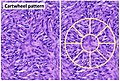 Histopathology of cartwheel pattern in dermatofibrosarcoma protuberans, annotated.jpg 2,701 × 1,830; 1.39 MB
-
 Histopathology of cartwheel pattern in dermatofibrosarcoma protuberans, original.jpg 2,048 × 1,532; 603 KB
-
 Histopathology of cholesterol clefts of a periapical cyst of the jaw.jpg 1,205 × 967; 593 KB
-
 Histopathology of chordoma, annotated.jpg 2,048 × 1,532; 656 KB
-
 Histopathology of chordoma, original.jpg 2,048 × 1,532; 410 KB
-
 Histopathology of corneal acute and chronic inflammation.jpg 1,229 × 1,491; 451 KB
-
 Histopathology of emphysema.jpg 1,104 × 1,136; 246 KB
-
 Histopathology of fibroadenoma with proliferating stroma.jpg 1,063 × 785; 338 KB
-
 Histopathology of fibroadenoma with small cell clusters.jpg 2,080 × 1,536; 931 KB
-
 Histopathology of ganglion cyst wall.jpg 1,721 × 1,536; 560 KB
-
 Histopathology of glioblastoma, high magnification, annotated.jpg 1,453 × 997; 368 KB
-
 Histopathology of glioblastoma, high magnification.jpg 2,048 × 1,532; 487 KB
-
 Histopathology of hyperplastic thyroid follicles in Graves' disease.jpg 2,048 × 1,532; 824 KB
-
 Histopathology of inverted urothelial papilloma, high magnification.jpg 1,530 × 1,522; 640 KB
-
 Histopathology of inverted urothelial papilloma.jpg 1,777 × 1,433; 835 KB
-
 Histopathology of irritation fibroma.jpg 3,140 × 2,048; 1.98 MB
-

-
 Histopathology of Kaposi's sarcoma.png 1,125 × 1,049; 1.84 MB
-
 Histopathology of lamina propria edema and hemorrhage in acute cholecystitis.jpg 2,048 × 1,532; 689 KB
-
 Histopathology of liposarcoma, annotated.jpg 1,945 × 1,473; 619 KB
-
 Histopathology of liposarcoma.jpg 2,048 × 1,532; 419 KB
-
 Histopathology of liver with cocci and granulomatous inflammation.jpg 2,048 × 1,532; 648 KB
-
 Histopathology of nasal squamous papilloma.jpg 1,063 × 749; 211 KB
-
 Histopathology of non-specific urothelial edema.jpg 2,048 × 1,532; 620 KB
-
 Histopathology of osteomyelitis.jpg 1,497 × 1,067; 326 KB
-
 Histopathology of osteosarcoma, high mag.jpg 2,048 × 1,532; 377 KB
-
 Histopathology of pancreatic adenocarcinoma with treatment effect.jpg 2,730 × 2,043; 770 KB
-
 Histopathology of pancreatic tissue in a mature cystic teratoma.jpg 2,048 × 1,532; 673 KB
-

-

-
 Histopathology of parathyroid adenoma.jpg 2,048 × 1,532; 524 KB
-
 Histopathology of parathyroid congestion.jpg 1,921 × 2,149; 1.51 MB
-
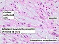
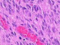
 Histopathology of pheochromocytoma (original).jpg 2,048 × 1,532; 639 KB
-
 Histopathology of pheochromocytoma.jpg 2,048 × 1,532; 804 KB
-
 Histopathology of pleomorphic adenoma (high magnification).jpg 2,045 × 2,633; 901 KB
-
 Histopathology of pleomorphic adenoma (low magnification).jpg 1,532 × 2,048; 820 KB
-
 Histopathology of pleomorphic adenoma.png 4,505 × 2,969; 14.05 MB
-
 Histopathology of seminoma, high magnification.jpg 2,048 × 1,532; 342 KB
-
 Histopathology of seminoma, intermediate magnification.jpg 2,048 × 1,532; 537 KB
-
 Histopathology of seminoma.png 3,071 × 2,122; 8.73 MB
-
 Histopathology of sinonasal acute inflammation, original.jpg 2,048 × 1,532; 377 KB
-
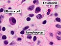 Histopathology of sinonasal inflammatory polyp with mixed inflammation, annotated.jpg 2,048 × 1,532; 440 KB
-
 Histopathology of sinonasal inflammatory polyp with mixed inflammation.jpg 2,048 × 1,532; 348 KB
-
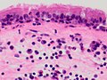 Histopathology of sinonasal inflammatory polyp.jpg 2,048 × 1,532; 393 KB
-
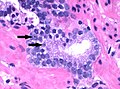 Histopathology of small acinar cell proliferation (annotated).jpg 2,080 × 1,536; 719 KB
-
 Histopathology of small acinar cell proliferation.jpg 2,080 × 1,536; 765 KB
-
 Histopathology of small bowel gangrene.jpg 1,160 × 857; 382 KB
-
 Histopathology of small cell carcinoma, annotated.png 2,048 × 1,532; 6.32 MB
-
 Histopathology of small cell carcinoma.jpg 2,048 × 1,532; 485 KB
-
 Histopathology of undifferentiated tumor cells.jpg 2,048 × 1,532; 431 KB
-
 Histopathology of vegetation of bacterial endocarditis.jpg 1,127 × 973; 341 KB
-
 Immunohistochemistry for CK19 in metastatic cholangiocarcinoma to the liver.jpg 2,048 × 1,532; 371 KB
-
 Immunohistochemistry of indeterminate expression of MSH-6.jpg 1,877 × 1,532; 385 KB
-
 Immunohistochemistry of prostein in metastatic prostate adenocarcinoma.jpg 2,048 × 1,532; 561 KB
-
 Immunohistochemistry stain for Melan-A in a metastatic melanoma to a lymph node.jpg 2,048 × 1,532; 355 KB
-
 Immunohistochemistry stain for SOX10 in a metastatic melanoma to a lymph node.jpg 2,048 × 1,532; 270 KB
-
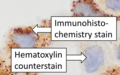 Immunohistochemistry stain versus counterstain.png 1,025 × 637; 683 KB
-
 Light microscopy with and without condenser.jpg 1,351 × 1,029; 401 KB
-
 Lipoblast features, annotated.png 2,048 × 1,532; 4.16 MB
-
 Lipoblast features.jpg 2,048 × 1,532; 442 KB
-
 Low- and high-grade urothelial carcinoma (original).jpg 2,691 × 1,997; 1.25 MB
-
 Low- and high-grade urothelial carcinoma.jpg 2,681 × 1,993; 1.45 MB
-
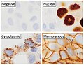 Main staining patterns on immunohistochemistry.jpg 2,137 × 1,657; 682 KB
-
 Marking a microscopy slide.jpg 2,752 × 3,067; 1.93 MB
-
 Measurement using transparent ruler in microscopy.jpg 4,681 × 2,697; 2.85 MB
-
 Measuring distance on a microscopy slide using a calibration slide.jpg 2,761 × 3,965; 1.48 MB
-
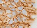 Membranous staining for E-cadherin on immunohistochemistry.jpg 2,048 × 1,532; 325 KB
-
 Micrograph of a calibration slide, showing the diameter of the field of view.jpg 3,029 × 3,033; 1.92 MB
-
 Micrograph of a melanophage.jpg 379 × 331; 39 KB
-
 Micrograph of marking a microscopy slide.jpg 4,897 × 2,441; 3.2 MB
-
 Negative staining on immunohistochemistry.jpg 2,048 × 1,532; 195 KB
-
 Osseous formation in a well-differentiated liposarcoma.jpg 2,048 × 1,532; 716 KB
-
 Pap stain of adenocarcinoma in peritoneal fluid.jpg 927 × 917; 139 KB
-
 Pap stain of adenocarcinoma in peritoneal fluid.png 1,149 × 975; 856 KB
-
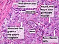
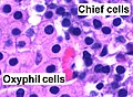 Parathyroid oxyphil and chief cells - annotated.jpg 911 × 665; 160 KB
-
 Parathyroid oxyphil and chief cells.jpg 2,080 × 1,536; 844 KB
-
 Pathology of avascular necrosis.jpg 2,093 × 1,893; 866 KB
-
 Photograph of marking a microscopy slide.jpg 3,053 × 2,085; 1.43 MB
-
 Plasmablast, Wright stain (hy).png 2,000 × 1,309; 1.65 MB
-
 Plasmablast, Wright stain, original.jpg 2,048 × 1,532; 260 KB
-
 Plasmablast, Wright stain.png 2,000 × 1,309; 2.01 MB
-
 Pleomorphic nuclei.jpg 878 × 690; 133 KB
-
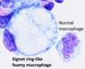 Signet ring-like foamy macrophage.png 1,521 × 1,237; 2.45 MB
-
 Signet-ring-cell-like foamy macrophage and normal macrophage.jpg 2,048 × 1,532; 293 KB
-
 Sinus histiocytosis (intermediate magnification).jpg 2,080 × 1,536; 1.13 MB
-
 Sinus histiocytosis (low magnification).jpg 2,080 × 1,536; 1.46 MB
-
 Subepicardial fibrosis (original).jpg 2,448 × 3,264; 2.29 MB
-
 Subepicardial fibrosis.jpg 1,031 × 1,352; 763 KB
-
 Thick blood film with Plasmodium falciparum rings and schizonts.png 506 × 354; 247 KB
-
 Trilineage hematopoiesis (original).jpg 2,080 × 1,536; 599 KB
-
 Trilineage hematopoiesis, annotated.jpg 773 × 595; 134 KB
-
 Whole slide image quality comparison.png 1,804 × 1,420; 2.06 MB
-
 Wright's staining of multiple myeloma, plasmablastic type.png 1,231 × 1,091; 1.6 MB


_stain_of_mixed_malignant_germ_cell_tumor_-_crop.png)
_stain_of_mixed_malignant_germ_cell_tumor.png)


.jpg)

.jpg)
.jpg)


.jpg)


.jpg)


.png)


.jpg)
.jpg)
.jpg)


.jpg)


