Category:Microscopic images of wood
Jump to navigation
Jump to search
Subcategories
This category has the following 11 subcategories, out of 11 total.
Media in category "Microscopic images of wood"
The following 58 files are in this category, out of 58 total.
-
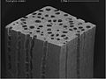
 20180422basswood19stack (41625441641).jpg 3,608 × 2,832; 3.25 MB
-
 Longitudinal section of tree trunk. Wellcome M0010773.jpg 2,433 × 4,335; 2.96 MB
-
 N. Grew, Cross-section of sumach,The anatomy of plants Wellcome L0016382.jpg 1,550 × 1,238; 755 KB
-
 Plant stem (250 26) Longitudal radial section - woody stem.jpg 3,752 × 2,401; 1.28 MB
-
 Plant stem (250 27) Longitudal radial section - woody stem.jpg 3,751 × 2,400; 1.83 MB
-

-

-
 Blue wooden button under a microscope 2.jpg 2,219 × 2,151; 830 KB
-
 Blue wooden button under a microscope.jpg 2,070 × 2,182; 1.01 MB
-
 Canale resinifero in sezione trasversale.jpg 1,492 × 1,600; 697 KB
-
 Dicotyledon-wood-cut.jpg 352 × 288; 25 KB
-
 Drevesina.jpg 1,488 × 1,249; 1.49 MB
-
 Ginkgo stem XS.jpg 817 × 600; 191 KB
-
 Large open ‘pores’ in a eucalypt.png 651 × 488; 232 KB
-
 Large open ‘pores’ in a Eucalyptus sieberi.jpg 651 × 488; 133 KB
-
 Meyers b8 s0668 b1.png 770 × 468; 179 KB
-
 Meyers b8 s0669 b1.png 753 × 498; 171 KB
-
 Microscopic image of Tectona grandis (Teak) 01.jpg 2,048 × 1,536; 584 KB
-
 Microscopy 10x pine mature wood.jpg 352 × 288; 41 KB
-
 Picture Natural History - No 318 - Section of Stem of Tree.png 653 × 596; 793 KB
-
 Plant stem (250 28) Longitudal radial section - woody stem.jpg 3,752 × 2,401; 1.96 MB
-
 Plant stem (255 12) Longitudal tangential section - woody stem.jpg 3,751 × 2,401; 1.84 MB
-
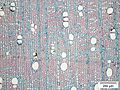
 Pore distribution in beech.jpg 563 × 423; 197 KB
-
 PSM V29 D450 Transverse sections of mill spalch and white cedar.jpg 2,012 × 1,034; 685 KB
-
 Quer digrep 1.jpg 1,600 × 1,200; 386 KB
-
 Querschnitt des Entadrophragma candollei.jpg 4,080 × 3,072; 3.71 MB
-
 Radial digrep 2.jpg 1,600 × 1,200; 243 KB
-
 Radialschnitt des Entadrophragma candollei.jpg 4,080 × 3,072; 3.19 MB
-
 Scary faces of wood 1.tif 1,280 × 1,280; 4.59 MB
-
 Scary faces of wood 2.tif 1,280 × 1,280; 4.26 MB
-
 Scary faces of wood 3.tif 1,280 × 1,280; 4.54 MB
-
 Scary faces of wood 4.tif 1,280 × 1,280; 4.18 MB
-
 Schima q200.jpg 4,080 × 3,072; 3.34 MB
-
 Schima rad200.jpg 4,080 × 3,072; 3.71 MB
-
 Schima tan200.jpg 4,080 × 3,072; 3.45 MB
-
 Spirostachys a. Radial (Crystals polar).jpg 1,600 × 1,200; 587 KB
-
 Spirostachys a. Radial (Crystals).jpg 1,600 × 1,200; 520 KB
-
 Spirostachys a. tangential 1.jpg 1,600 × 1,200; 501 KB
-
 Spirostachys africana radial.jpg 1,600 × 1,200; 496 KB
-
 Tangential digrep1.jpg 1,600 × 1,200; 251 KB
-
 Tangentialschnitt des Entadrophragma candollei.jpg 4,080 × 3,072; 3.51 MB
-
 Transverse section Spirostachys africana.JPG 1,600 × 1,200; 529 KB
-
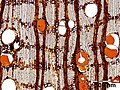
 Typical diffuse-porous wood species.jpg 2,337 × 1,449; 1.28 MB
-
 Whole set of Scary faces of wood with scale bar.png 16,188 × 4,000; 84.21 MB
-
 Whole set of Scary faces of wood.png 16,188 × 4,000; 84.22 MB
-
 Wood fibers 100X.jpg 3,264 × 2,448; 1.81 MB
-
 Wood fibers 40X.jpg 3,264 × 2,448; 2.5 MB
-

-

-
 Woody Dicot Stem.jpg 967 × 982; 687 KB
-
 XylemInToothpick.JPG 1,024 × 717; 183 KB
-
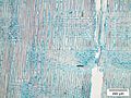
 Zellarten im Laubholz.jpg 2,823 × 4,136; 1.2 MB
-
 Zellarten im Nadelholz.jpg 2,823 × 2,155; 626 KB
-
 Zweig eines Ahorn im Querschnitt.jpg 800 × 640; 109 KB
-
 Окаймлённая пора ( похожа на глаз дракона).jpg 963 × 691; 388 KB
-
 Поперечный срез стебля Ginkgo biloba.jpg 7,874 × 7,874; 24.52 MB
-
 Секреторная клетка Гинкго билоба.JPG 5,472 × 3,648; 5.78 MB
-
 Срез ветви дерева под микроскопом.JPG 2,204 × 3,920; 1.28 MB
.jpg)


_Longitudal_radial_section_-_woody_stem.jpg)
_Longitudal_radial_section_-_woody_stem.jpg)
_(17550764793).jpg)
_(17985165779).jpg)


_01.jpg)


_Longitudal_radial_section_-_woody_stem.jpg)
_Longitudal_tangential_section_-_woody_stem.jpg)


.jpg)
.jpg)


_(20783612906).jpg)
_(20800431012).jpg)


.jpg)


