Category:Human cerebellum
Jump to navigation
Jump to search
Subcategories
This category has the following 22 subcategories, out of 22 total.
- SVG human cerebellum (18 F)
A
- Anterior lobe of cerebellum (29 F)
- Arbor vitae (anatomy) (44 F)
C
- Cerebellar cortex (18 F)
- Cerebellar fossa (19 F)
- Cerebellar hemisphere (20 F)
- Cerebellar lobules (11 F)
- Cerebellar neurons (2 F)
- Cerebellar tonsil (15 F)
- Cerebellar vermis (60 F)
F
- Flocculonodular lobe (20 F)
H
P
- Posterior lobe of cerebellum (24 F)
- Human purkinje cells (1 F)
T
Media in category "Human cerebellum"
The following 124 files are in this category, out of 124 total.
-
1312 CerebellumN.jpg 875 × 1,080; 348 KB
-
1543, Andreas Vesalius' Fabrica, Base Of The Brain.jpg 1,202 × 1,346; 1.11 MB
-
1543,AndreasVesalius'Fabrica,BaseOfTheBrain.png 292 × 308; 164 KB
-
1612 Cerebellar Peduncles-02.jpg 1,421 × 1,083; 648 KB
-
1613 Major Regions of the Cerebellum-02.jpg 2,248 × 1,167; 976 KB
-
201405 cerebellem.png 400 × 400; 62 KB
-
4 spill 2 knowing-neurons2.jpg 838 × 1,024; 84 KB
-
Afférences du cervelet - version simplifiée.png 782 × 684; 28 KB
-
Blausen 0115 BrainStructures.png 1,600 × 1,429; 1.68 MB
-
BodyParts3D FJ3834 FJ3876 Cerebellum.stl 5,120 × 2,880; 2.27 MB
-
Boucle de Gullain-morallet - néocervelet.png 798 × 430; 17 KB
-
Brain-cerebellum.png 201 × 169; 38 KB
-
CerebCircuit.png 388 × 377; 23 KB
-
Cerebellar blood-flow.png 790 × 934; 2.82 MB
-
Cerebellar Circuit grayscale German.png 1,143 × 1,215; 89 KB
-
Cerebellar fossa by Sanjoy Sanyal.webm 44 s, 718 × 404; 4.51 MB
-
Cerebellar glomerulus-vi.tif 885 × 735; 661 KB
-
Cerebellar glomerulus.tif 885 × 735; 662 KB
-
Cerebellum 4.jpg 960 × 720; 118 KB
-
Cerebellum animation small.gif 150 × 150; 443 KB
-
Cerebellum cross-section, without labels (colorized).png 951 × 579; 509 KB
-
Cerebellum cross-section, without labels.png 951 × 579; 317 KB
-
Cerebellum Dissection Video 1 - FULL - Sanjoy Sanyal 1 of 3.webm 4 min 47 s, 640 × 360; 45.65 MB
-
Cerebellum Dissection Video 1 - FULL - Sanjoy Sanyal 2 of 3.webm 6 min 23 s, 640 × 360; 57.47 MB
-
Cerebellum Dissection Video 1 - FULL - Sanjoy Sanyal 3 of 3.webm 5 min 15 s, 640 × 360; 49.01 MB
-
Cerebellum Dissection Video 2 - Sagittal - Sanjoy Sanyal.webm 5 min 48 s, 638 × 360; 55.39 MB
-
Cerebellum Dissection Video 3 - Axial View - Sanjoy Sanyal.webm 5 min 6 s, 638 × 360; 43.73 MB
-
Cerebellum Einteilung.png 914 × 637; 52 KB
-
Cerebellum NIH.png 360 × 262; 114 KB
-
Cerebellum sag.jpg 300 × 223; 24 KB
-
Cerebellum sagittal planes.png 675 × 518; 253 KB
-
Cerebellum small.gif 151 × 150; 527 KB
-
Cerebellum somatotopie - PT.jpg 845 × 607; 266 KB
-
Cerebellum.png 800 × 455; 325 KB
-
CerebellumDiv zh.png 914 × 637; 59 KB
-
CerebellumDiv-es.png 1,000 × 697; 157 KB
-
CerebellumDiv-vi.png 940 × 672; 66 KB
-
CerebellumDiv.png 914 × 637; 67 KB
-
CerebellumDivRO.png 914 × 637; 52 KB
-
CerebellumRegions.jpg 309 × 263; 77 KB
-
Diseases of the nervous system (1908) (14592660687).jpg 2,096 × 1,664; 1.29 MB
-
Diseases of the nervous system (1910) (14586285529).jpg 2,104 × 1,664; 571 KB
-
-
-
Gray677.png 400 × 370; 25 KB
-
Gray702.png 600 × 319; 73 KB
-
Gray703 (Flocculus).png 600 × 350; 215 KB
-
Gray703.png 600 × 350; 59 KB
-
Gray704.png 600 × 416; 59 KB
-
Gray705.png 500 × 374; 42 KB
-
Gray706-vi.png 622 × 638; 162 KB
-
Gray706.png 550 × 598; 56 KB
-
Gray707 zh.png 400 × 319; 95 KB
-
Gray707-vi.png 400 × 319; 100 KB
-
Gray707.png 400 × 319; 39 KB
-
Gray708.png 600 × 385; 78 KB
-
Gray709.png 500 × 442; 49 KB
-
Gray745.png 550 × 447; 62 KB
-
Gray768.png 500 × 408; 37 KB
-
Horizontal sections of fetal brain.jpg 960 × 720; 123 KB
-
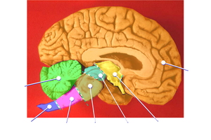
Human brain midsagittal view description.JPG 423 × 374; 30 KB
-
Human brainstem Sagittal view.jpg 490 × 360; 146 KB
-
Human cerebellum anterior view description.JPG 344 × 200; 16 KB
-
Human cerebellum anterior view.JPG 344 × 200; 14 KB
-
Human cerebellum posterior view description.JPG 345 × 211; 18 KB
-
Human cerebellum posterior view.JPG 345 × 211; 16 KB
-
Illu cerebrum lobes.jpg 371 × 274; 34 KB
-
Illu diencephalon-ar.jpg 350 × 263; 13 KB
-
Illu diencephalon.jpg 350 × 263; 17 KB
-
Image from page 632 of "Human physiology" (1856) (14762204386).jpg 1,518 × 1,598; 285 KB
-
Kleinhirn-es.png 1,600 × 1,099; 252 KB
-
Kleinhirn.png 1,577 × 1,083; 124 KB
-
Lawrence 1960 18.1.png 2,268 × 1,692; 1.02 MB
-
Lawrence 1960 18.4.png 2,376 × 1,268; 849 KB
-
Lawrence 1960 2.43.png 1,996 × 2,588; 1 MB
-
Lawrence 1960 2.44.png 1,976 × 2,412; 863 KB
-
Lawrence 1960 2.45.png 2,212 × 1,776; 932 KB
-
Lawrence 1960 2.46.png 2,212 × 932; 396 KB
-
Location of the foramina of the fourth ventricle.jpg 650 × 462; 56 KB
-
Model of Cerebellar Perceptron.jpg 464 × 379; 27 KB
-
MRI image of cerebellum.png 623 × 1,606; 663 KB
-
Neuron counts of cerebral cortex and cerebellum.png 358 × 341; 102 KB
-
Néocervelet.png 826 × 535; 23 KB
-
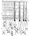
Parallel-fibers.png 389 × 465; 64 KB
-
Porbeagle shark brain.png 394 × 130; 11 KB
-
Posterior view of the human cerebellum.png 677 × 475; 165 KB
-
PSM V26 D760 Upper surface of the cerebellum.jpg 1,247 × 915; 295 KB
-
PSM V26 D761 Inferior surface of the cerebellum.jpg 1,273 × 872; 310 KB
-
PSM V42 D021 Outer surface of the cerebellum.jpg 1,669 × 1,307; 177 KB
-
Pääsukese saba.jpg 1,280 × 720; 559 KB
-
Reichert "Der bau des menschlichen...", 1859; cerebellum Wellcome L0016337.jpg 1,218 × 1,570; 703 KB
-
Rhombencephalon.jpg 960 × 720; 70 KB
-
Sagittal scan of fetal head Ecografia Dr. Wolfgang Moroder.theora.ogv 13 s, 976 × 735; 4.43 MB
-
Sheep brain.jpg 768 × 1,024; 222 KB
-
Slide2AST-ar.jpg 960 × 720; 174 KB
-
Slide2AST.JPG 960 × 720; 81 KB
-
Slide2BRA-ar.jpg 960 × 720; 200 KB
-
Slide2BRA.JPG 960 × 720; 103 KB
-
Slide2PIT.JPG 960 × 720; 97 KB
-
Slide2SEER.JPG 960 × 720; 89 KB
-
Slide3AST.JPG 960 × 720; 92 KB
-
Slide3EER.JPG 960 × 720; 89 KB
-
Slide3PIT.JPG 960 × 720; 105 KB
-
Slide4SER.JPG 960 × 720; 112 KB
-
Sobo 1909 624 ar.png 3,060 × 2,247; 5.38 MB
-
Sobo 1909 624.png 3,060 × 2,247; 19.71 MB
-
Sobo 1909 642.png 1,063 × 1,019; 3.11 MB
-
Sobo 1909 647.png 1,168 × 1,165; 3.9 MB
-
Sobo 1909 648.png 1,063 × 1,048; 3.19 MB
-
Sobo 1909 653.png 1,004 × 512; 1.47 MB
-
Sobo 1909 654.png 1,007 × 507; 1.46 MB
-
Sobo 1909 655.png 1,022 × 539; 1.58 MB
-
Sobo 1909 656.png 867 × 498; 1.24 MB
-
Sobo 1909 657.png 904 × 513; 1.33 MB
-
Sobo 1909 658.png 951 × 579; 1.58 MB
-
Spinocervelet.png 698 × 593; 17 KB
-
Thomas-fig67-p124.png 279 × 244; 9 KB
-
Thomas-fig68,69-p127.png 412 × 307; 97 KB
-
Ultrasound Scan ND 0117084539 0852090.png 800 × 600; 145 KB
-
Ultrasound Scan ND 0117084539 0852140.png 800 × 600; 147 KB
-
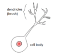
Unipolar brush cell.GIF 305 × 293; 5 KB
-
Vestibulocervelet.png 939 × 534; 21 KB
-
Треугольник Рейля.jpg 297 × 185; 41 KB
-
Углубление Рейля.jpg 297 × 207; 40 KB


