Category:Human brain (sagittal section)
Jump to navigation
Jump to search
anatomical plane dividing the body into left and right | |||||
| Upload media | |||||
| Subclass of |
| ||||
|---|---|---|---|---|---|
| |||||
Subcategories
This category has the following 2 subcategories, out of 2 total.
Media in category "Human brain (sagittal section)"
The following 149 files are in this category, out of 149 total.
-
4167134294 d0cf029385 bTêteCoupe.jpg 2,451 × 3,050; 487 KB
-
7 Tesla MRI of the ex vivo human brain at 100 micron resolution (100 micron MRI acquired FA25 sagittal).webm 1 min 25 s, 1,760 × 1,280; 89.55 MB
-
A text-book of human physiology (1906) (14770191352).jpg 1,716 × 1,346; 196 KB
-
Acute Effects of Ecstasy.gif 720 × 540; 169 KB
-
AddictionDependence-de.png 480 × 374; 224 KB
-
AddictionDependence-de.xcf 480 × 374; 998 KB
-
Addictiondependence1-es.png 480 × 374; 55 KB
-
Addictiondependence1-German.png 480 × 374; 262 KB
-
Addictiondependence1.png 480 × 374; 137 KB
-
Adverse Effects of Ecstasy.gif 720 × 540; 174 KB
-
Aktivitaethinten.jpg 220 × 239; 24 KB
-
Anatomy and physiology LS dog's brain.JPG 612 × 367; 46 KB
-
Basic structures of the brain highlighted.png 945 × 763; 267 KB
-
Blausen 0104 Brain x-secs SectionalPlanes-ar.jpg 2,250 × 1,600; 463 KB
-
Blausen 0104 Brain x-secs SectionalPlanes-Arabic-YM.png 2,250 × 1,600; 2.21 MB
-
Blausen 0104 Brain x-secs SectionalPlanes.png 2,250 × 1,600; 1.85 MB
-
Bourgery & Jacob-cs04cl.jpg 1,944 × 2,272; 2.55 MB
-
Brain (PSF).jpg 807 × 622; 89 KB
-
Brain Anatomy (Sagittal) ku.png 809 × 618; 444 KB
-
Brain Anatomy (Sagittal).png 1,024 × 768; 618 KB
-
Brain Areas Affected by Ecstasy.gif 720 × 540; 168 KB
-
Brain bulbar region.PNG 295 × 299; 51 KB
-
Brain chrischan 300.gif 300 × 300; 756 KB
-
Brain chrischan 600.gif 600 × 600; 2.56 MB
-
Brain chrischan thalamus.jpg 860 × 860; 71 KB
-
Brain chrischan.jpg 860 × 860; 70 KB
-
Brain Maksymilian Rose.PNG 779 × 479; 169 KB
-
Brain midsagital view.png 1,024 × 768; 262 KB
-
Brain sagittal.png 936 × 890; 142 KB
-
Brain surgery (1893) (14781134142).jpg 1,380 × 1,838; 875 KB
-
Brain-isolines.png 641 × 565; 101 KB
-
Bulbe rachidien4.jpg 609 × 611; 73 KB
-
Cerebral vascular territories midline.jpg 1,024 × 999; 127 KB
-
Cingulate region of human brain.jpg 336 × 259; 20 KB
-
Corpuis callosum.png 624 × 624; 302 KB
-
Cortical midline structures.png 634 × 532; 324 KB
-
Cortical spreading depression-AR.gif 295 × 299; 29 KB
-
Cortical spreading depression-IT.gif 295 × 299; 28 KB
-
Cortical spreading depression.gif 295 × 299; 29 KB
-
Coupe sagit noyau accumbens5.jpg 1,035 × 825; 101 KB
-
Coupe section du tronc cérébral.png 927 × 1,024; 195 KB
-
Die Frau als Hausärztin (1911) 012 Das Gehirn.png 349 × 509; 271 KB
-
Diseases of the nervous system (1910) (14749919176).jpg 2,200 × 1,316; 367 KB
-
DTI-sagittal-xyzrgb.jpg 1,021 × 1,125; 64 KB
-
EB1911 Brain Fig. 11-Gyri and Sulci on Mesial aspect.jpg 682 × 393; 45 KB
-
Engraving; section of brain, by Capieux, 1792. Wellcome L0007162.jpg 1,500 × 1,298; 710 KB
-
Faisceau pedicule.jpg 600 × 600; 48 KB
-
FlechsigSaggital4.jpg 500 × 443; 223 KB
-
GarpenBrain.jpg 1,280 × 1,024; 677 KB
-
Gray 720-emphasizing-corpus-callosum.png 600 × 526; 1.21 MB
-
Gray1180-ar.png 672 × 375; 165 KB
-
Gray1180.png 672 × 375; 45 KB
-
Gray518.png 500 × 288; 27 KB
-
Gray568.png 600 × 363; 50 KB
-
Gray715.png 600 × 443; 158 KB
-
Gray720.png 600 × 526; 126 KB
-
Gray745.png 550 × 447; 62 KB
-
Gray751.png 500 × 307; 26 KB
-
Gray768.png 500 × 408; 37 KB
-
Gyrus cinguli.png 800 × 467; 116 KB
-
Human brain anatomical axes alterations.jpg 1,000 × 1,000; 243 KB
-
Human Brain Dissected.jpg 2,521 × 2,046; 979 KB
-
Human brain left dissected midsagittal view description 2.JPG 701 × 488; 50 KB
-
Human brain left midsagitttal view closeup description 3.JPG 701 × 490; 58 KB
-
Human brain midsagittal cut color.png 1,294 × 861; 1.17 MB
-
Human brain midsagittal cut color2.png 958 × 720; 1.14 MB
-
Human brain midsagittal cut description.JPG 701 × 486; 50 KB
-
Human brain midsagittal cut.JPG 701 × 486; 45 KB
-
Human brain midsagittal view description.JPG 423 × 374; 30 KB
-
Human brain right dissected lateral view description.JPG 653 × 413; 40 KB
-
Human Brain Sagittal Section.JPG 1,024 × 768; 231 KB
-
Human brain, sagittal section 1.jpg 3,073 × 2,858; 1.51 MB
-
Human brain.jpg 843 × 1,011; 209 KB
-
Human Brain2.png 200 × 280; 43 KB
-
Human brainstem Sagittal view.jpg 490 × 360; 146 KB
-
Human telencephalon Rose 1938.PNG 937 × 635; 412 KB
-
Hypothalamus.jpg 236 × 248; 11 KB
-
Inferior colliculus - sagittal cut.jpg 817 × 581; 244 KB
-
Labeledbrain.jpg 860 × 860; 78 KB
-
LocationOfHypothalamus.jpg 350 × 250; 21 KB
-
Meynert1885.PNG 1,400 × 867; 1.1 MB
-
Microcephaly.png 648 × 237; 157 KB
-
Mozek obratlovců – oblasti.png 471 × 678; 289 KB
-
MRI brain sagittal section.jpg 645 × 702; 100 KB
-
Mri brain side view-emphasizing-corpus-callosum.png 600 × 600; 1.38 MB
-
Mri brain side view.jpg 900 × 900; 225 KB
-
Mri head 3dani 1 small bionerd.gif 200 × 243; 734 KB
-
MRI head side.jpg 256 × 256; 10 KB
-
MRI.ogv 5.5 s, 240 × 256; 1.34 MB
-
MRI.png 1,512 × 1,056; 6.77 MB
-
Mrt big.jpg 820 × 754; 192 KB
-
Nervous and mental diseases (1911) (14775794734).jpg 2,088 × 1,280; 252 KB
-
Nervous and mental diseases (1919) (14758523716).jpg 2,088 × 1,276; 199 KB
-
Neurologische Reaktion bei Trauma, Stress und Tremor Response.pdf 1,239 × 1,754; 69 KB
-
Neurones noradrenergiques.png 649 × 450; 167 KB
-
Noradrenergic neurons ar.png 649 × 450; 166 KB
-
Nucleus accumbens.jpg 332 × 289; 20 KB
-
Núcleos del rafé 01.png 509 × 667; 147 KB
-
Onufrowicz balkendefekte.PNG 883 × 531; 61 KB
-
Operative surgery (1905) (14597206190).jpg 2,912 × 2,182; 1.13 MB
-
PET1.jpg 422 × 348; 16 KB
-
Pituitary gland representation.PNG 261 × 236; 8 KB
-
Pituitary gland.png 468 × 332; 210 KB
-
Possible effects of nicotine on the developing human brain (cropped).jpg 1,818 × 1,660; 1.02 MB
-
Possible effects of nicotine on the developing human brain.jpg 2,838 × 2,843; 941 KB
-
Primary central nervous system B-cell non-Hodgkin lymphoma.jpg 729 × 729; 193 KB
-
Principles of Psychology (James) v1 p36b.png 1,520 × 890; 824 KB
-
PSM V01 D357 Human encephalon.jpg 974 × 762; 147 KB
-
PSM V46 D168 Mesial view of the human brain.jpg 1,765 × 1,480; 470 KB
-
Ptsd-brain.png 441 × 421; 183 KB
-
Recklinghausen2.jpg 509 × 443; 25 KB
-
Sagittal brain slice, UB Museum of Neuroanatomy, Buffalo, New York.jpg 1,280 × 1,920; 1.19 MB
-
Sagittal MRI scan of brain of patient with Chiari malformation.jpg 1,200 × 1,344; 212 KB
-
Sagittale-insula-Heschl.png 579 × 420; 140 KB
-
Serotonin nerve pathways in the brain.gif 720 × 540; 172 KB
-
Side View of the Brain.png 1,275 × 1,650; 2.09 MB
-
Slide2ZEN.JPG 960 × 720; 115 KB
-
Slide3ZEN.JPG 960 × 720; 120 KB
-
Smegenys(brain).png 960 × 720; 261 KB
-
Sobo 1909 624 ar.png 3,060 × 2,247; 5.38 MB
-
Sobo 1909 624.png 3,060 × 2,247; 19.71 MB
-
Sobo 1909 634.png 1,089 × 703; 2.19 MB
-
Steele-olszewski-richardson disease.jpg 705 × 591; 95 KB
-
Structural MRI animation.ogv 5.6 s, 256 × 256; 1.76 MB
-
The brain as an organ of mind (1896) (14760990156).jpg 1,504 × 1,228; 585 KB
-
Tumor Germinoma PinealGland2.JPG 927 × 861; 61 KB
-
Tumor Germinoma Suprasellar2.JPG 807 × 830; 55 KB
-
Tumor PrimaryCNSLymphoma T1Saggital.JPG 607 × 751; 35 KB
-
VBM1.jpg 426 × 391; 18 KB
-
Ventricular system of the brain BG language tags.jpg 1,650 × 1,275; 298 KB
-
Vertebrate-brain-regions small hy.png 300 × 432; 161 KB
-
Vertebrate-brain-regions small.png 300 × 432; 147 KB
-
Vertebrate-brain-regions-he.png 471 × 678; 218 KB
-
Vertebrate-brain-regions.png 471 × 678; 90 KB
-
Vue-hemisphere-droit-cerveau-humain.jpg 766 × 486; 76 KB
-
Vuyvem600.png 600 × 600; 139 KB
-
Webysther 635864498758569254 - Imagem por ressonância magnética.png 480 × 480; 170 KB
-
Žmogaus smegenų struktūra.png 519 × 409; 287 KB
-
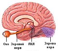
Зоровий шлях.jpg 250 × 218; 45 KB
-
Магнитно-резонансная томография.jpg 960 × 1,280; 97 KB
-
Позвоночные — мозг — области.png 471 × 678; 291 KB
-
۱۴۰۰Brain.png 689 × 391; 198 KB
-
মানব মানব মস্তিষ্ক.jpg 600 × 352; 33 KB


