Category:Human brain (inferior view)
Jump to navigation
Jump to search
Subcategories
This category has the following 2 subcategories, out of 2 total.
Media in category "Human brain (inferior view)"
The following 67 files are in this category, out of 67 total.
-
"Traite complet de l'anatomie...",Foville, 1844 Wellcome L0019135.jpg 3,763 × 4,947; 4.58 MB
-
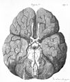
1543, Andreas Vesalius' Fabrica, Base Of The Brain.jpg 1,202 × 1,346; 1.11 MB
-
1543,AndreasVesalius'Fabrica,BaseOfTheBrain.png 292 × 308; 164 KB
-
A9. tb meningitis.jpg 480 × 640; 23 KB
-
Amentia Plate IA.jpg 1,799 × 2,738; 865 KB
-
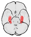
Amyg.png 189 × 230; 22 KB
-
Anatomical model of human brain, Wellcome L0010008.jpg 1,474 × 1,186; 469 KB
-
Anatomical model of human brain, Wellcome L0010009.jpg 1,440 × 1,216; 467 KB
-
ArterieCervello.jpg 635 × 585; 101 KB
-
Arteries beneath brain Gray closer-ar.jpg 635 × 585; 310 KB
-
Arteries beneath brain Gray closer.jpg 635 × 585; 104 KB
-
Arteries beneath brain.png 851 × 681; 207 KB
-
Autopsy brain.jpg 1,600 × 1,059; 328 KB
-
Bernard - La science expérimentale fig-2.jpg 1,668 × 1,684; 753 KB
-
Brain human normal inferior view with labels ar.png 424 × 505; 362 KB
-
Brain; dissection showing the base of the brain. Coloured li Wellcome V0008405.jpg 2,153 × 3,289; 3.14 MB
-
Brain; dissection showing the base of the brain. Coloured li Wellcome V0008408.jpg 2,151 × 3,377; 3.42 MB
-
Brain; dissection showing the base of the brain. Watercolour Wellcome V0008415ER.jpg 1,485 × 1,951; 1.82 MB
-
Brodmann area 20.png 256 × 192; 38 KB
-
Brodmann area 38.png 256 × 192; 33 KB
-
Circle of Willis Wellcome M0016877.jpg 3,071 × 3,595; 3.91 MB
-
Circle of Willis.jpg 995 × 1,097; 696 KB
-
Constudproc.png 763 × 865; 31 KB
-
Facies ventralis cerebri.jpg 1,834 × 2,068; 629 KB
-
FrontalCaptsBasal.png 932 × 537; 243 KB
-
Fusiform Gyrus and its Sulci on 3D-printed brain, inferior view.png 2,441 × 2,893; 4.19 MB
-
Fusiform Gyrus on 3D-printed brain, inferior view.png 2,441 × 2,893; 4.34 MB
-
Gray724.png 600 × 588; 73 KB
-
Gray729.png 300 × 366; 20 KB
-
Gray748.png 500 × 510; 66 KB
-
Hippocampus.png 231 × 274; 39 KB
-
Hippocampus.svg 254 × 295; 258 KB
-
Human base of brain blood supply description.JPG 501 × 540; 42 KB
-
Human base of brain blood supply.JPG 501 × 540; 38 KB
-
Human brain anterior-inferior view description.JPG 330 × 475; 31 KB
-
Human brain anterior-inferior view.JPG 330 × 475; 28 KB
-
Human brain inferior view description 2.JPG 373 × 467; 36 KB
-
Human brain inferior view description.JPG 373 × 466; 37 KB
-
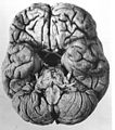
Human brain inferior view.JPG 373 × 467; 34 KB
-
Human brainstem anterior view 2 description.JPG 346 × 487; 35 KB
-
Human brainstem anterior view blood supply description.JPG 335 × 466; 30 KB
-
Human brainstem anterior view blood supply.JPG 335 × 466; 30 KB
-
Human brainstem anterior view description 2.JPG 347 × 485; 31 KB
-
Human brainstem anterior view description.JPG 347 × 485; 31 KB
-
Human brainstem anterior view.JPG 347 × 485; 27 KB
-
Human brainstem blood supply description.JPG 331 × 468; 36 KB
-
Inferior human brain coloured.svg 320 × 392; 202 KB
-
J. M. Charcot, Diseases of the nervous syste Wellcome L0029908.jpg 4,310 × 2,602; 4.57 MB
-
Location of Midbrain in inferior view.png 373 × 467; 307 KB
-
Optic nerve pair & two brain hemispheres.jpg 252 × 405; 21 KB
-
Optic processing human brain.jpg 501 × 700; 137 KB
-
Plate 16, 'The anatomy of humane bodies...' Wellcome L0063587.jpg 3,633 × 6,424; 7.91 MB
-
PSM V26 D758 Under surface of the human brain.jpg 1,089 × 1,332; 381 KB
-
Skull inner surface.jpg 2,409 × 3,134; 5.27 MB
-
Slide13ee.JPG 960 × 720; 138 KB
-
Slide2MIR.JPG 960 × 720; 115 KB
-
Slide2PIT.JPG 960 × 720; 97 KB
-
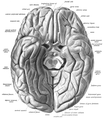
Sobo 1909 623 ar.png 2,404 × 2,652; 5.62 MB
-
Sobo 1909 623.png 2,404 × 2,652; 18.27 MB
-
Sobo 1909 629.png 991 × 1,015; 98 KB
-
Sobo 1909 630.png 1,077 × 1,239; 3.83 MB
-
Structures of brain.jpg 935 × 648; 104 KB
-
The Brain Wellcome L0008287.jpg 1,546 × 1,216; 964 KB
-
The brain, in right profile with the glossopharyngeal and va Wellcome V0010423.jpg 2,290 × 3,270; 3.81 MB
-
Trigeminal ganglion.jpg 960 × 720; 130 KB
-
Tuberculous leptomeningitis.jpg 1,682 × 2,215; 1.35 MB
-
মানব মানব মস্তিষ্ক.jpg 600 × 352; 33 KB


