Category:Human anatomy from CNX Anatomy & Physiology Textbook
Appearance
Subcategories
This category has the following 4 subcategories, out of 4 total.
Media in category "Human anatomy from CNX Anatomy & Physiology Textbook"
The following 200 files are in this category, out of 325 total.
(previous page) (next page)-
01 01ab Gross and Microscopic Anatomy.jpg 825 × 351; 243 KB
-
01 16 X-ray of Hand (cropped).jpg 469 × 469; 110 KB
-
01 16 X-ray of Hand.jpg 750 × 881; 207 KB
-
0910 Oateoarthritis Hip B.png 712 × 791; 274 KB
-
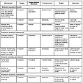
1125 Muscles in Facial Expression.jpg 1,019 × 1,338; 513 KB
-
1126 Understand A Muscle from the Latin.jpg 1,933 × 758; 411 KB
-
1127 Muscles for Tongue Movement Swallowing and Speech.jpg 2,088 × 2,717; 1.76 MB
-
1128 Muscles of the Perineum Common to Men and Women.jpg 1,944 × 1,905; 948 KB
-
1129 Muscles that Moves the Humerus.jpg 970 × 1,090; 497 KB
-
1130 Muscles that Move the Forearm.jpg 1,971 × 1,983; 1.04 MB
-
1131 Muscles that Moves the Wrist Hands and Fingers.jpg 2,263 × 3,063; 2.04 MB
-
1132 Gluteal Region Muscles that Move the Femur.jpg 2,275 × 3,425; 2.17 MB
-
1133 Thigh Muscles that Moves the Femur Tibia and Fibula.jpg 1,971 × 2,746; 1.5 MB
-
1134 Muscles that Moves the Feet and Toes.jpg 2,239 × 3,075; 1.63 MB
-
1135 Intrinsic Muscles in the Foot.jpg 1,958 × 2,753; 1.45 MB
-
1201 Overview of Nervous System esp.jpg 841 × 760; 218 KB
-
1201 Overview of Nervous System nl01.jpg 877 × 867; 167 KB
-
1201 Overview of Nervous System zh.jpg 841 × 760; 166 KB
-
1201 Overview of Nervous System.jpg 841 × 760; 319 KB
-
1202 White and Gray Matter.jpg 975 × 714; 641 KB
-
1204 Optic Nerve vs Optic Tract.jpg 787 × 615; 215 KB
-
1305 CerebrumN.jpg 1,119 × 521; 216 KB
-
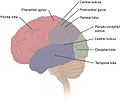
1306 Lobes of Cerebral CortexN.jpg 733 × 621; 160 KB
-
1307 Brodmann Areas.jpg 2,353 × 1,394; 1.38 MB
-
1308 Frontal Section Basal Nuclei.jpg 684 × 535; 164 KB
-
1309 Basal Nuclei Connections.jpg 500 × 496; 103 KB
-
1310 Diencephalon.jpg 869 × 689; 218 KB
-
1311 Brain Stem-es.png 1,000 × 756; 443 KB
-
1311 Brain Stem.jpg 906 × 685; 208 KB
-
1312 CerebellumN.jpg 875 × 1,080; 348 KB
-
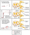
1313 Spinal Cord Cross Section.jpg 1,642 × 1,883; 1.6 MB
-
1315 Brain Sinuses.jpg 1,090 × 826; 537 KB
-
1317 CFS Circulation -ru -v1.jpg 1,057 × 693; 316 KB
-
1317 CFS Circulation -ru -v2.jpg 1,057 × 693; 317 KB
-
1317 CFS Circulation.jpg 1,065 × 693; 373 KB
-
1320 The Cranial Nerves pl.jpg 962 × 491; 240 KB
-
1320 The Cranial Nerves.jpg 784 × 491; 230 KB
-
1321 Spinal Nerve Plexuses.jpg 752 × 1,263; 415 KB
-
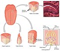
1402 The Tongue esp.jpg 2,229 × 1,875; 1,015 KB
-
1402 The Tongue.jpg 2,229 × 1,875; 1.29 MB
-
1404 The Structures of the Ear.jpg 1,721 × 1,063; 652 KB
-
1406 Cochlea esp.jpg 2,063 × 996; 559 KB
-
1406 Cochlea.jpg 2,063 × 996; 699 KB
-
1407 The Hair Cell.jpg 2,188 × 971; 803 KB
-
1408 Frequency Coding in The Cochlea.jpg 2,213 × 1,421; 812 KB
-
1409 Maculae and Equilibrium.jpg 2,388 × 1,254; 1.05 MB
-
1410 Equilibrium and Semicircular Canals.jpg 2,256 × 1,112; 809 KB
-
1411 Eye in The Orbit.jpg 1,742 × 1,140; 808 KB
-
1413 Structure of the Eye.jpg 2,175 × 1,242; 1.06 MB
-
1417 Ascending Pathways of Spinal Cord.jpg 2,271 × 2,325; 1.18 MB
-
1424 Visual Streams.jpg 2,146 × 1,033; 592 KB
-
1426 Corticospinal Pathway-es.png 1,200 × 2,627; 909 KB
-
1426 Corticospinal Pathway.jpg 1,163 × 2,546; 851 KB
-
1427 Cochlea Micrograph.jpg 1,621 × 992; 868 KB
-
1501 Connections of the Sympathetic Nervous System-es.png 1,040 × 1,515; 700 KB
-
1501 Connections of the Sympathetic Nervous System.jpg 970 × 1,427; 657 KB
-
1502 Symphatetic Connections and the Ganglia.jpg 1,950 × 2,321; 1.3 MB
-
1503 Connections of the Parasympathetic Nervous System.jpg 1,967 × 3,021; 676 KB
-
1510 Fiber Tracts of the Central Autonomic System.jpg 1,663 × 1,488; 845 KB
-
1511 The Limbic Lobe-ar.png 1,950 × 1,111; 592 KB
-
1511 The Limbic Lobe-vi.jpg 1,950 × 1,111; 354 KB
-
1511 The Limbic Lobe.jpg 1,950 × 1,111; 617 KB
-
1512 Connections to Heart.jpg 1,813 × 1,570; 567 KB
-
1601 Anatomical Underpinnings of the Neurological Exam-02.jpg 1,256 × 1,500; 370 KB
-
1603 Brodmann Areas-02.jpg 2,333 × 1,369; 1.39 MB
-
1604 Types of Cortical Areas-02 (ja).jpg 1,915 × 989; 184 KB
-
1604 Types of Cortical Areas-02.jpg 1,915 × 989; 568 KB
-
1605 Brocas and Wernickes Areas-02 (ja).jpg 1,463 × 992; 103 KB
-
1605 Brocas and Wernickes Areas-02-ar.jpg 1,463 × 992; 282 KB
-
1605 Brocas and Wernickes Areas-02.jpg 1,463 × 992; 331 KB
-
1611 Dermatomes-02.jpg 1,242 × 2,279; 936 KB
-
1612 Cerebellar Peduncles-02.jpg 1,421 × 1,083; 648 KB
-
1613 Major Regions of the Cerebellum-02.jpg 2,248 × 1,167; 976 KB
-
1615 Locations Spinal Fiber Tracts.jpg 1,475 × 1,483; 474 KB
-
1801 The Endocrine System-ar.jpg 928 × 831; 127 KB
-
1801 The Endocrine System.jpg 928 × 831; 282 KB
-
1806 The Hypothalamus-Pituitary Complex esp.jpg 2,154 × 1,252; 500 KB
-
1806 The Hypothalamus-Pituitary Complex.jpg 1,077 × 626; 248 KB
-
1807 The Posterior Pituitary Complex esp.jpg 2,196 × 1,774; 633 KB
-
1807 The Posterior Pituitary Complex.jpg 1,098 × 887; 335 KB
-
1808 The Anterior Pituitary Complex esp.jpg 2,308 × 1,992; 711 KB
-
1808 The Anterior Pituitary Complex.jpg 1,154 × 996; 372 KB
-
1811 The Thyroid Gland.jpg 737 × 1,410; 542 KB
-
1813 A Classic Negative Feedback Loop.jpg 1,122 × 1,097; 484 KB
-
1814 The Parathyroid Glands.jpg 1,117 × 408; 347 KB
-
1817 The Role of Parathyroid Hormone in Maintaining Blood Calcium Homeostasis.jpg 1,186 × 1,425; 803 KB
-
1818 The Adrenal Glands-ar.jpg 1,102 × 316; 106 KB
-
1818 The Adrenal Glands-es.jpg 1,300 × 373; 318 KB
-
1818 The Adrenal Glands.jpg 1,102 × 316; 250 KB
-
1820 The Pancreas.jpg 1,092 × 551; 423 KB
-
2000 Human Heart Photo.jpg 1,671 × 1,179; 1.03 MB
-
2001 Heart Position in ThoraxN.jpg 2,177 × 1,974; 1.22 MB
-
201 Elements of the Human Body-01-body.jpg 791 × 1,274; 126 KB
-
201 Elements of the Human Body-01-body.png 791 × 1,274; 137 KB
-
201 Elements of the Human Body-01-es.png 2,300 × 1,294; 395 KB
-
201 Elements of the Human Body-01.jpg 2,276 × 1,280; 599 KB
-
2010 Chordae Tendinae Papillary Muscles.jpg 1,606 × 1,112; 869 KB
-
2032 Automatic Innervation.fr.jpg 1,110 × 2,071; 413 KB
-
2032 Automatic Innervation.jpg 1,110 × 2,071; 803 KB
-
2110 Pulse Sites.jpg 1,063 × 1,633; 358 KB
-
2119 Pulmonary Circuit.jpg 1,507 × 868; 665 KB
-
2125 Thoracic Abdominal Arteries Chart.jpg 1,971 × 2,796; 1.32 MB
-
2126 Iliac Artery Branches Chart.jpg 1,821 × 1,496; 688 KB
-
2128 Thoracic Upper Limb Arteries Chart.jpg 2,263 × 1,933; 1.05 MB
-
2130 Lower Limb Arteries Chart.jpg 2,233 × 2,271; 804 KB
-
2135 Veins Draining into Superior Vena Cava Chart.jpg 2,279 × 2,296; 1.09 MB
-
2137 Lower Limb Veins Chart.jpg 1,838 × 2,000; 576 KB
-
2138 Hepatic Portal Vein System.jpg 1,583 × 1,021; 583 KB
-
2140 FlowChart Veins into VenaCava.jpg 2,263 × 2,471; 923 KB
-
2141 CircSyst vs OtherSystemsN.jpg 1,870 × 2,257; 1.74 MB
-
2201 Anatomy of the Lymphatic System.jpg 1,115 × 1,181; 678 KB
-
2203 Lymphatic Trunks and Ducts System.jpg 870 × 678; 267 KB
-
2206 The Location Structure and Histology of the Thymus.jpg 1,084 × 760; 502 KB
-
2208 Spleen.jpg 1,036 × 979; 506 KB
-
2209 Location and Histology of Tonsils.jpg 910 × 1,173; 540 KB
-
2301 Major Respiratory Organs.jpg 1,933 × 1,625; 804 KB
-
2302 External Nose.jpg 1,936 × 1,728; 546 KB
-
2303 Anatomy of Nose-Pharynx-Mouth-Larynx.jpg 1,917 × 1,600; 926 KB
-
2305 Divisions of the Pharynx.jpg 1,956 × 1,496; 400 KB
-
2306 The Larynx.jpg 1,814 × 1,673; 649 KB
-
2307 Cartilages of the Larynx.jpg 1,938 × 1,133; 481 KB
-
2308 The Trachea.jpg 1,952 × 1,430; 1.06 MB
-
2308a The Trachea.jpg 1,459 × 2,159; 761 KB
-
2310 Structures of the Respiratory Zone-a.jpg 1,496 × 1,782; 809 KB
-
2312 Gross Anatomy of the Lungs.jpg 1,896 × 1,304; 611 KB
-
2313 The Lung Pleurea.jpg 1,925 × 1,450; 845 KB
-
2327 Respiratory Centers of the Brain esp.jpg 1,917 × 2,471; 1.05 MB
-
2327 Respiratory Centers of the Brain.jpg 1,917 × 2,471; 1.08 MB
-
2401 Components of the Digestive System.jpg 936 × 1,185; 363 KB
-
2403 The PeritoneumN.jpg 826 × 589; 292 KB
-
2406 Structures of the Mouth.jpg 1,075 × 1,028; 388 KB
-
2407 Tongue.jpg 626 × 557; 209 KB
-
2408 Salivary Glands.jpg 586 × 542; 191 KB
-
2410 Permanent and Deciduous TeethN.jpg 660 × 1,110; 336 KB
-
2411 Pharynx.jpg 792 × 927; 231 KB
-
2412 The Esophagus.jpg 619 × 824; 164 KB
-
2413 DeglutitionN.jpg 1,017 × 575; 242 KB
-
2414 Stomach.jpg 1,122 × 741; 466 KB
-
2415 Histology of StomachN.jpg 1,214 × 568; 415 KB
-
2417 Small IntestineN.jpg 839 × 552; 166 KB
-
2418 Histology Small IntestinesN esp.jpg 2,158 × 1,648; 1.17 MB
-
2418 Histology Small IntestinesN.jpg 1,079 × 824; 540 KB
-
2420 Large Intestine.jpg 633 × 427; 163 KB
-
2421 Histology of the Large IntestineN.jpg 1,085 × 923; 608 KB
-
2422 Accessory Organs.jpg 805 × 1,002; 224 KB
-
2424 Exocrine and Endocrine Pancreas FR.jpg 2,635 × 2,329; 643 KB
-
2424 Exocrine and Endocrine Pancreas-it.jpg 637 × 827; 367 KB
-
2424 Exocrine and Endocrine Pancreas.jpg 637 × 827; 441 KB
-
2425 Gallbladder-es.png 918 × 661; 238 KB
-
2425 Gallbladder-tr.png 787 × 641; 187 KB
-
2425 Gallbladder.jpg 787 × 641; 212 KB
-
2429 Digestion of Proteins (Physiology).jpg 641 × 803; 224 KB
-
2433 Teniae Coli Haustra Epiploic Appendage.jpg 636 × 699; 213 KB
-
2602 Female Urethra.jpg 1,729 × 1,253; 907 KB
-
2603 Male Urethra N.jpg 1,854 × 1,492; 1.07 MB
-
2604 Nerves Innervating the Urinary SystemN-fr.jpg 1,738 × 1,265; 601 KB
-
2604 Nerves Innervating the Urinary SystemN.jpg 1,738 × 1,265; 327 KB
-
2605 The Bladder zh.jpg 1,962 × 1,202; 984 KB
-
2605 The Bladder-es.png 2,000 × 1,225; 525 KB
-
2605 The Bladder.jpg 1,962 × 1,202; 1.09 MB
-
2608 Kidney Position in Abdomen.jpg 1,630 × 1,204; 600 KB
-
2610 The Kidney.jpg 1,975 × 1,129; 844 KB
-
2612 Blood Flow in the Kidneys.jpg 1,971 × 1,565; 1.12 MB
-
2911 Photo of Placenta-02.jpg 1,661 × 1,264; 1.26 MB
-
2921 Neonatal Circulatory System.jpg 1,950 × 1,651; 1.04 MB
-
3d CT scan animation.gif 250 × 250; 994 KB
-
401 Types of Tissue.jpg 1,070 × 1,135; 633 KB
-
413 Types of Membranes.jpg 769 × 1,123; 355 KB
-
5 ligaments of interosseous membrane of forearm.png 717 × 1,254; 288 KB
-
507 Nails.jpg 1,206 × 364; 211 KB
-
516 Aging.jpg 975 × 689; 348 KB
-
601 Bone Classification Cleaned.png 692 × 755; 257 KB
-
601 Bone Classification.jpg 1,012 × 1,173; 368 KB
-
602 Bone Markings.jpg 992 × 1,065; 347 KB
-
603 Anatomy of Long Bone zh.jpg 708 × 1,156; 152 KB
-
603 Anatomy of Long Bone.jpg 708 × 1,156; 257 KB
-
609 Body Supply to the Bone.jpg 582 × 810; 220 KB
-
610 Feature Pagets Disease.jpg 598 × 617; 214 KB
-
610 Feature Pagets Disease.png 1,205 × 1,243; 175 KB
-
615 Age and Bone Mass-ar.jpg 860 × 553; 236 KB
-
615 Age and Bone Mass.jpg 860 × 553; 218 KB
-
618 Bones Protect Brain.jpg 621 × 805; 227 KB
-
623 Epiphyseal Plate-Line.jpg 790 × 625; 157 KB
-
700 Lateral View of Skull-01.jpg 1,963 × 1,806; 2.65 MB
-
701 Axial Skeleton-01.jpg 2,406 × 2,447; 1.35 MB
-
702 Newborn Skull-01.jpg 2,383 × 803; 433 KB
-
703 Parts of Skull-01.jpg 1,664 × 1,378; 459 KB
-
704 Skull-01.jpg 2,437 × 2,062; 1.2 MB
-
705 Lateral View of Skull-01-ar.svg 735 × 504; 395 KB
-
705 Lateral View of Skull-01.jpg 2,450 × 1,681; 1.06 MB
-
705 Lateral View of Skull-01.svg 735 × 504; 136 KB
-
706 Sagittal Section of Skull-01.jpg 2,454 × 1,696; 1.23 MB
-
707 Superior-Inferior View of Skull Base-01.jpg 2,283 × 3,096; 1.82 MB
-
708 Temporal Bone.jpg 774 × 555; 177 KB
-
709 Sphenoid Bone.jpg 634 × 820; 244 KB
-
710 Ethmoid Bone.jpg 825 × 637; 179 KB
-
711 Maxilla.jpg 784 × 601; 166 KB
-
712 Hyoid Bone - ja.jpg 545 × 847; 155 KB
-
712 Hyoid Bone.jpg 545 × 847; 180 KB
-
713 Bones Forming Orbit.jpg 965 × 457; 182 KB


