Category:Cell biology from CNX Anatomy & Physiology Textbook
Jump to navigation
Jump to search
Media in category "Cell biology from CNX Anatomy & Physiology Textbook"
The following 62 files are in this category, out of 62 total.
-
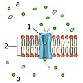
0303 Lipid Bilayer With Various Components labeled.jpg 875 × 400; 311 KB
-
0303 Lipid Bilayer With Various Components-ar.jpg 5,833 × 2,667; 5.01 MB
-
0303 Lipid Bilayer With Various Components.jpg 875 × 400; 389 KB
-
0306 Facilitated Diffusion Carrier Protein labeled.jpg 728 × 384; 151 KB
-
0306 Facilitated Diffusion Carrier Protein.jpg 781 × 384; 158 KB
-
0306 Facilitated Diffusion Channel Protein labeled.jpg 530 × 532; 107 KB
-
0306 Facilitated Diffusion Channel Protein.jpg 563 × 532; 115 KB
-
0306 Facilitated Diffusion.jpg 781 × 987; 338 KB
-
0308 Sodium Potassium Pump labeled.jpg 1,016 × 500; 266 KB
-
0308 Sodium Potassium Pump.jpg 1,052 × 500; 327 KB
-
0309 Phagocytosis cleaned.png 282 × 425; 63 KB
-
0309 Phagocytosis.png 375 × 485; 67 KB
-
0309 Pinocytosis cleaned.png 369 × 412; 66 KB
-
0309 Pinocytosis.png 366 × 485; 70 KB
-
0309 RME cleaned.png 350 × 453; 66 KB
-
0309 RME.png 381 × 485; 78 KB
-
0309 Three Forms of Endocytosis cleaned.jpg 1,008 × 460; 183 KB
-
0309 Three Forms of Endocytosis.jpg 1,124 × 513; 259 KB
-
0310 Exocytosis cleaned.png 544 × 432; 100 KB
-
0310 Exocytosis.jpg 544 × 612; 125 KB
-
0311 Pancreatic Cells Micrograph labeled.jpg 694 × 390; 163 KB
-
0311 Pancreatic Cells Micrograph.jpg 822 × 390; 219 KB
-
0312 Animal Cell and Components.jpg 813 × 645; 388 KB
-
0313 Endoplasmic Reticulum a en.png 604 × 362; 230 KB
-
0313 Endoplasmic Reticulum a labeled.png 604 × 362; 253 KB
-
0313 Endoplasmic Reticulum b en.png 467 × 363; 190 KB
-
0313 Endoplasmic Reticulum b labeled.png 371 × 363; 196 KB
-
0313 Endoplasmic Reticulum c en.png 733 × 398; 245 KB
-
0313 Endoplasmic Reticulum c labeled.png 604 × 398; 250 KB
-
0313 Endoplasmic Reticulum.jpg 1,102 × 892; 488 KB
-
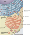
0314 Golgi Apparatus a ar.png 579 × 708; 357 KB
-
0314 Golgi Apparatus a en.png 579 × 708; 368 KB
-
0314 Golgi Apparatus a ru.png 605 × 708; 436 KB
-
0314 Golgi Apparatus b en.png 521 × 435; 234 KB
-
0314 Golgi Apparatus.jpg 1,124 × 754; 494 KB
-
0315 Mitochondrion new.jpg 2,233 × 991; 987 KB
-
0316 Peroxisome.jpg 541 × 443; 157 KB
-
0317 Cytoskeletal Components.jpg 1,109 × 554; 483 KB
-
0318 Nucleus.jpg 777 × 599; 355 KB
-
0320 RBC Extruding Nucleus Micrograph.jpg 1,117 × 384; 518 KB
-
0321 DNA Macrostructure.jpg 743 × 584; 227 KB
-
0323 DNA Replication.jpg 819 × 506; 291 KB
-
0326 Splicing.jpg 580 × 667; 134 KB
-
0327 Translation.jpg 542 × 1,025; 206 KB
-
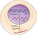
0328 Transcription-translation Summary.jpg 548 × 525; 169 KB
-
0329 Cell Cycle.jpg 531 × 433; 116 KB
-
0330 Homologous Pair of Chromosomes.jpg 482 × 411; 135 KB
-
0331 Stages of Mitosis and Cytokinesis.jpg 1,118 × 861; 691 KB
-
0332 Cell Cycle With Cyclins and Checkpoints.jpg 666 × 583; 172 KB
-
0338 RNA Polymerase Binding.jpg 1,118 × 245; 109 KB
-
1215 Cell Membrane Channels.jpg 1,143 × 544; 405 KB
-
1216 Ligand-gated Channels.jpg 1,176 × 611; 606 KB
-
1217 Mechanically-gated Channels-02.jpg 1,133 × 580; 523 KB
-
1218 Voltage-gated Channels.jpg 1,102 × 548; 476 KB
-
1219 Leakage Channels.jpg 1,119 × 525; 474 KB
-
1220 Resting Membrane Potential.jpg 1,119 × 541; 474 KB
-
1803 Binding of Lipid-Soluble Hormones.jpg 937 × 621; 357 KB
-
1804 Binding of Water-Soluble Hormones.jpg 965 × 731; 441 KB
-
2508 The Electron Transport Chain.jpg 1,975 × 1,358; 931 KB
-
2705 Sodium Potassium Pump.jpg 1,079 × 499; 327 KB
-
2706 Facilitated Diffusion.jpg 777 × 377; 199 KB
-
402 Types of Cell Junctions new.jpg 4,597 × 5,366; 2.02 MB


