File:Post-Embryonic-Nerve-Associated-Precursors-to-Adult-Pigment-Cells-Genetic-Requirements-and-Dynamics-pgen.1002044.s007.ogv
Jump to navigation
Jump to search
Size of this JPG preview of this OGG file: 600 × 600 pixels. Other resolutions: 240 × 240 pixels | 480 × 480 pixels | 642 × 642 pixels.
Original file (Ogg Theora video file, length 12 s, 642 × 642 pixels, 449 kbps, file size: 630 KB)
Captions
Captions
Add a one-line explanation of what this file represents
Summary[edit]
| Description |
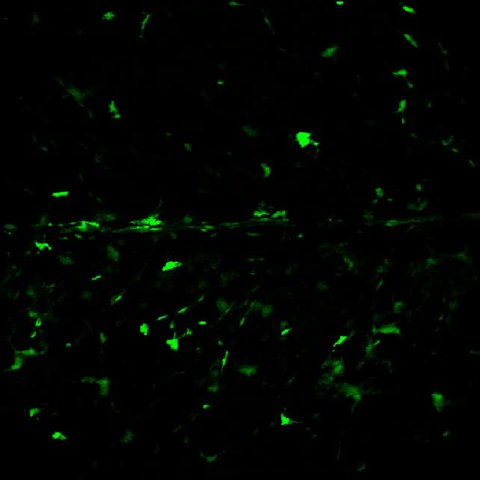
English: mitfa::GFP+ cells in a living wild-type larva. Images were collected in sagittal planes from the skin to the midline of the trunk. Image rotation reveals extra-hypodermal cells within the myotomes, that typically expressed mitfa::GFP at levels lower than in hypodermal cells. |
||
| Date | |||
| Source | Video S1 from Budi E, Patterson L, Parichy D (2011). "Post-Embryonic Nerve-Associated Precursors to Adult Pigment Cells: Genetic Requirements and Dynamics of Morphogenesis and Differentiation". PLOS Genetics. DOI:10.1371/journal.pgen.1002044. PMID 21625562. PMC: 3098192. | ||
| Author | Budi E, Patterson L, Parichy D | ||
| Permission (Reusing this file) |
|
||
| Provenance |
|
File history
Click on a date/time to view the file as it appeared at that time.
| Date/Time | Thumbnail | Dimensions | User | Comment | |
|---|---|---|---|---|---|
| current | 22:32, 5 June 2013 | 12 s, 642 × 642 (630 KB) | Open Access Media Importer Bot (talk | contribs) | Automatically uploaded media file from Open Access source. Please report problems or suggestions here. |
You cannot overwrite this file.
File usage on Commons
There are no pages that use this file.
Transcode status
Update transcode statusMetadata
Categories:
- Videos of developmental biology
- Molecular development
- Videos of cell migration
- Videos of stem cells
- Adult stem cells
- Stem cell niche
- Cell differentiation
- Cell fate determination
- Organism development
- Gene function
- Videos of mutations
- Cell lines
- Cell shape
- Videos of cell biology
- Developmental gene expression regulation
- ErbB-3 receptor