File:Parasite130116-1-olm Entamoeba gingivalis microscopy.tif
Jump to navigation
Jump to search

Size of this JPG preview of this TIF file: 800 × 597 pixels. Other resolutions: 320 × 239 pixels | 640 × 478 pixels | 1,024 × 764 pixels | 1,280 × 956 pixels | 1,436 × 1,072 pixels.
Original file (1,436 × 1,072 pixels, file size: 1,010 KB, MIME type: image/tiff)
Captions
Captions
Add a one-line explanation of what this file represents
Summary[edit]
| Description |
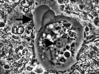
English: Entamoeba gingivalis - Microscopic diagnosis
Supplemental material from article. In periodontitis or healthy sites, periodontal material including dental plaque was sampled with a probe. The sample was saliva-mounted and immediately observed by phase-contrast microscopy. Amoebae were detected by their producing one lobose pseudopodium at a time, with dark intracellular vacuoles and one nucleus containing a central karyosome and peripheral chromatin. |
| Date | |
| Source | Bonner, M., Amard, V., Bar-Pinatel, C., Charpentier, F., Chatard, J.-M., Desmuyck, Y., Ihler, S., Rochet, J.-P., Roux de La Tribouille, V., Saladin, L., Verdy, M., Gironès, N., Fresno, M. & Santi-Rocca, J. 2014: Detection of the amoeba Entamoeba gingivalis in periodontal pockets. Parasite, 21, 30. doi:10.1051/parasite/2014029 |
| Author | Mark Bonner, Véronique Amard, Charlotte Bar-Pinatel, Frédéric Charpentier, Jean-Michel Chatard, Yvan Desmuyck, Serge Ihler, Jean-Pierre Rochet, Véronique Roux de La Tribouille, Luc Saladin, Marion Verdy, Núria Gironès, Manuel Fresno and Julien Santi-Rocca |
Licensing[edit]
This file is licensed under the Creative Commons Attribution 4.0 International license.
|
 This file was published in the scientific journal Parasite. Their website states that all content of the journal including and after 2013 is published under the Creative Commons Attribution 4.0 license.
|
File history
Click on a date/time to view the file as it appeared at that time.
| Date/Time | Thumbnail | Dimensions | User | Comment | |
|---|---|---|---|---|---|
| current | 15:55, 7 July 2014 |  | 1,436 × 1,072 (1,010 KB) | Jeanloujustine (talk | contribs) | User created page with UploadWizard |
You cannot overwrite this file.
File usage on Commons
The following page uses this file:
File usage on other wikis
The following other wikis use this file:
- Usage on ar.wikipedia.org
- Usage on arz.wikipedia.org
- Usage on az.wikipedia.org
- Usage on de.wikipedia.org
- Usage on eml.wikipedia.org
- Usage on en.wikipedia.org
- Usage on fa.wikipedia.org
- Usage on fr.wikipedia.org
- Usage on gl.wikipedia.org
- Usage on id.wikipedia.org
- Usage on it.wikipedia.org
- Usage on pl.wikipedia.org
- Usage on pt.wikipedia.org
- Usage on ru.wikipedia.org
- Usage on species.wikimedia.org
- Usage on tr.wikipedia.org
- Usage on www.wikidata.org