File:E15.5 mouse embryonic testes.tif
Jump to navigation
Jump to search

Size of this JPG preview of this TIF file: 796 × 599 pixels. Other resolutions: 319 × 240 pixels | 638 × 480 pixels | 1,020 × 768 pixels | 1,280 × 964 pixels | 1,360 × 1,024 pixels.
Original file (1,360 × 1,024 pixels, file size: 4.01 MB, MIME type: image/tiff)
Captions
Captions
Add a one-line explanation of what this file represents
Summary[edit]
| Description |
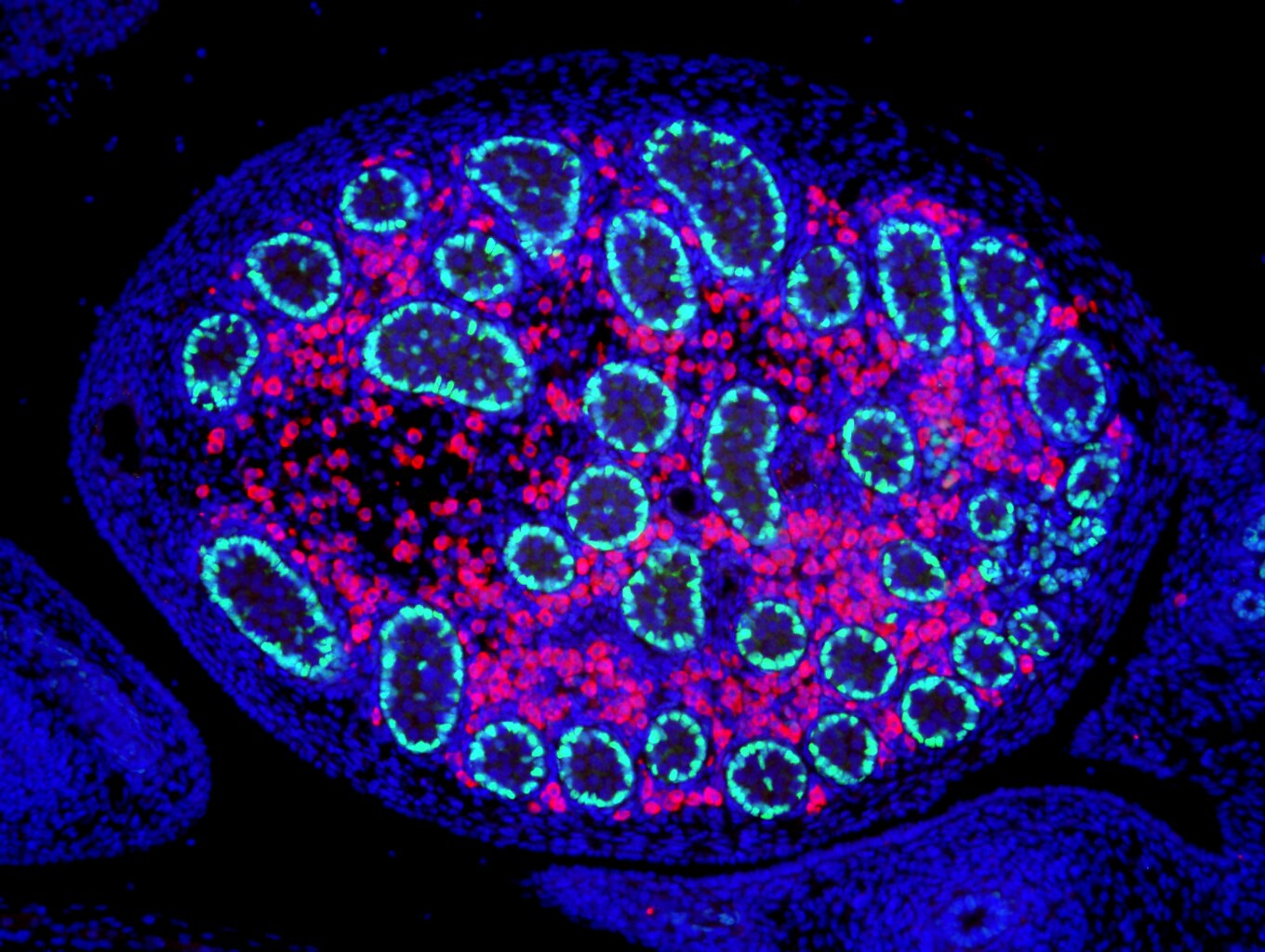
English: A paraffin section of E15.5 wildtype mouse testis fluorescently stained with antibodies detecting the Sertoli cell marker SOX9 (green) and the Leydig cell marker 3beta-HSD (red). Nuclei (blue) are stained with DAPI. Sertoli cells are found within the tubular-like testes cords and play multiple roles in testes cell differentiation and morphogenesis. On the other hand, Leydig cells populate the interstitial compartment and mediate male steroidogenesis.Hudson Institute of Medical Research, Melbourne, Australia. |
| Date | |
| Source | Own work |
| Author | Adbird81 |
Licensing[edit]
I, the copyright holder of this work, hereby publish it under the following license:
This file is licensed under the Creative Commons Attribution 4.0 International license.
- You are free:
- to share – to copy, distribute and transmit the work
- to remix – to adapt the work
- Under the following conditions:
- attribution – You must give appropriate credit, provide a link to the license, and indicate if changes were made. You may do so in any reasonable manner, but not in any way that suggests the licensor endorses you or your use.
| This image was uploaded as part of Wiki Science Competition 2017. |
File history
Click on a date/time to view the file as it appeared at that time.
| Date/Time | Thumbnail | Dimensions | User | Comment | |
|---|---|---|---|---|---|
| current | 01:17, 6 December 2017 |  | 1,360 × 1,024 (4.01 MB) | Adbird81 (talk | contribs) | User created page with UploadWizard |
You cannot overwrite this file.
File usage on Commons
The following 31 pages use this file:
- UV light
- Commons:Wiki Science Competition 2017
- Commons:Wiki Science Competition 2017/Images
- Commons:Wiki Science Competition 2017/Images/ar
- Commons:Wiki Science Competition 2017/Images/da
- Commons:Wiki Science Competition 2017/Images/de
- Commons:Wiki Science Competition 2017/Images/en
- Commons:Wiki Science Competition 2017/Images/fr
- Commons:Wiki Science Competition 2017/Images/it
- Commons:Wiki Science Competition 2017/Images/mk
- Commons:Wiki Science Competition 2017/Images/pl
- Commons:Wiki Science Competition 2017/Winners/Australia
- Commons:Wiki Science Competition 2017/Winners/Microscopy images
- Commons:Wiki Science Competition 2017/ar
- Commons:Wiki Science Competition 2017/da
- Commons:Wiki Science Competition 2017/de
- Commons:Wiki Science Competition 2017/en
- Commons:Wiki Science Competition 2017/es
- Commons:Wiki Science Competition 2017/fr
- Commons:Wiki Science Competition 2017/it
- Commons:Wiki Science Competition 2017/mk
- Commons:Wiki Science Competition 2017/pl
- Commons:Wiki Science Competition 2017/pt-br
- Commons:Wiki Science Competition 2017/ru
- Commons:Wiki Science Competition 2017/th
- Commons:Wiki Science Competition 2017/tr
- Commons:Wiki Science Competition 2017/vi
- Commons:Wiki Science Competition 2019 in Russia
- Category:General category images from Russian Science Photo Competition 2020
- Category:Wiki Science Competition 2019 in Russia
- Category:Wiki Science Competition 2020 in Russia
File usage on other wikis
The following other wikis use this file: