File:Dynamic-3D-Cell-Rearrangements-Guided-by-a-Fibronectin-Matrix-Underlie-Somitogenesis-pone.0007429.s005.ogv
Jump to navigation
Jump to search
Size of this JPG preview of this OGG file: 600 × 600 pixels. Other resolutions: 240 × 240 pixels | 480 × 480 pixels.
Original file (Ogg Theora video file, length 25 s, 800 × 800 pixels, 1.57 Mbps, file size: 4.74 MB)
Captions
Captions
Add a one-line explanation of what this file represents
Summary[edit]
| Description |
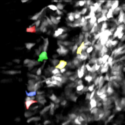
English: Multiphoton 4D image sequence of GFP expressing-cells in chick embryo PSM showing cell movements during somite formation. First segment of the video shows a portion of the PSM of a chick embryo (rostral is upwards, medial is to the left). In the beginning only one somite is formed, but at the end of 6 hours four new somites have formed. After zooming into a single somite the details of individual cell movements are shown. Several different morphogenetic stereotypical movements are identified in cells with different colors. Blue represents elongation of a medial cell, red cell accretion in the rostral and caudal walls, green egression, and yellow mesenchymal elongation and intercalation in the lateral wall. In the end, a zoom of a forming intersomitic cleft shows the continuous extension and retraction of pseudopodia. For more details refer to Figure 2 of the manuscript. |
||
| Date | |||
| Source | Video S3 from Martins G, Rifes P, Amândio R, Rodrigues G, Palmeirim I, Thorsteinsdóttir S (2009). "Dynamic 3D Cell Rearrangements Guided by a Fibronectin Matrix Underlie Somitogenesis". PLOS ONE. DOI:10.1371/journal.pone.0007429. PMID 19829711. PMC: 2759537. | ||
| Author | Martins G, Rifes P, Amândio R, Rodrigues G, Palmeirim I, Thorsteinsdóttir S | ||
| Permission (Reusing this file) |
|
||
| Provenance |
|
File history
Click on a date/time to view the file as it appeared at that time.
| Date/Time | Thumbnail | Dimensions | User | Comment | |
|---|---|---|---|---|---|
| current | 00:47, 14 November 2012 | 25 s, 800 × 800 (4.74 MB) | Open Access Media Importer Bot (talk | contribs) | Automatically uploaded media file from Open Access source. Please report problems or suggestions here. |
You cannot overwrite this file.
File usage on Commons
There are no pages that use this file.