File:A-versatile-pipeline-for-the-multi-scale-digital-reconstruction-and-quantitative-analysis-of-3D-elife-11214-media1.ogv
Jump to navigation
Jump to search
Size of this JPG preview of this OGG file: 733 × 600 pixels. Other resolutions: 293 × 240 pixels | 587 × 480 pixels | 880 × 720 pixels.
Original file (Ogg Theora video file, length 10 s, 880 × 720 pixels, 2.65 Mbps, file size: 3.29 MB)
Captions
Captions
Add a one-line explanation of what this file represents
Summary[edit]
| Description |
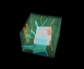
English: 3D image visualization of a multi-resolution geometrical model of liver tissue. A set of six low-resolution (1.0 μm × 1.0 μm × 2.0 μm per voxel) and two high-resolution tissue sections (0.3 μm × 0.3 μm × 0.3 μm per voxel) were used. Central veins are shown in light blue, portal veins in orange and high-resolution cubes in grey.
DOI: https://dx.doi.org/10.7554/eLife.11214.012 |
||
| Date | |||
| Source | Video 1. from Morales-Navarrete H, Segovia-Miranda F, Klukowski P, Meyer K, Nonaka H, Marsico G, Chernykh M, Kalaidzidis A, Zerial M, Kalaidzidis Y (2015). "A versatile pipeline for the multi-scale digital reconstruction and quantitative analysis of 3D tissue architecture". eLife. DOI:10.7554/eLife.11214. PMID 26673893. PMC: 4764584. | ||
| Author | Morales-Navarrete H, Segovia-Miranda F, Klukowski P, Meyer K, Nonaka H, Marsico G, Chernykh M, Kalaidzidis A, Zerial M, Kalaidzidis Y | ||
| Permission (Reusing this file) |
This file is licensed under the Creative Commons Attribution 4.0 International license.
|
||
| Provenance |
|
File history
Click on a date/time to view the file as it appeared at that time.
| Date/Time | Thumbnail | Dimensions | User | Comment | |
|---|---|---|---|---|---|
| current | 04:46, 29 February 2016 | 10 s, 880 × 720 (3.29 MB) | Open Access Media Importer Bot (talk | contribs) | Automatically uploaded media file from Open Access source. Please report problems or suggestions here. |
You cannot overwrite this file.
File usage on Commons
The following page uses this file:
Transcode status
Update transcode statusFile usage on other wikis
The following other wikis use this file:
- Usage on outreach.wikimedia.org