File:A-gene-trap-transposon-eliminates-haematopoietic-expression-of-zebrafish-Gfi1aa-but-does-not-mmc7.ogv
Jump to navigation
Jump to search
Size of this JPG preview of this OGG file: 800 × 450 pixels. Other resolutions: 320 × 180 pixels | 640 × 360 pixels | 960 × 540 pixels.
Original file (Ogg multiplexed audio/video file, Theora/Vorbis, length 32 s, 960 × 540 pixels, 845 kbps overall, file size: 3.19 MB)
Captions
Captions
Add a one-line explanation of what this file represents
Summary
[edit]| Description |
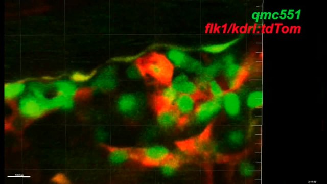
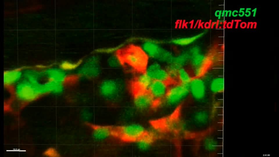
English: Movie 6 : qmc551: GFP-positive haematopoietic cells seed perivascular niches in the caudal haematopoietic tissue at 2 dpf and patrol the head tissues at 6 dpf. This movie is related to Figure 3. (A) Timelapse confocal microscopy on qmc551;flk1: tdTom double transgenic embryos starting from 48 hpf showing 2.0 µm thick optical slices through the caudal haematopoietic tissue. Here, GFP+ haematopoietic cells undergo dynamic interactions with tdTom+ ECs. One of the cells is highlighted with a yellow circle. Please note that ECs in the ventral wall of the caudal artery co-express the two transgenes. Anterior is to the left, dorsal is up. The embryo shown was immobilized in 1% low melting point agarose. (B) A 6 dpf qmc551 transgenic embryo was anesthetized and placed in a tiny depression in agarose under a fluorescent Nikon SMZ1500 dissection microscope using a Nikon DS-5Mc/DS-U1 camera setup. From minute 5 on, pictures of the head region of the embryo (facing right) were taken manually every 3 min. Images were imported into Photoshop. In Photoshop, the pictures were moved and rotated to correct for the drifting movement of the embryo under the microscope. Furthermore, annotations were added. All pictures were then imported into iMovie to generate the final video. Individual frames are also shown in Figure 3O. |
||
| Date | |||
| Source | Video file from Thambyrajah R, Ucanok D, Jalali M, Hough Y, Wilkinson R, McMahon K, Moore C, Gering M (2016). "A gene trap transposon eliminates haematopoietic expression of zebrafish Gfi1aa, but does not interfere with haematopoiesis". Developmental Biology. DOI:10.1016/j.ydbio.2016.07.010. PMID 27432513. PMC: 5003831. | ||
| Author | Thambyrajah R, Ucanok D, Jalali M, Hough Y, Wilkinson R, McMahon K, Moore C, Gering M | ||
| Permission (Reusing this file) |
This file is licensed under the Creative Commons Attribution 4.0 International license.
|
||
| Provenance |
|
File history
Click on a date/time to view the file as it appeared at that time.
| Date/Time | Thumbnail | Dimensions | User | Comment | |
|---|---|---|---|---|---|
| current | 20:02, 29 October 2016 | 32 s, 960 × 540 (3.19 MB) | Open Access Media Importer Bot (talk | contribs) | Automatically uploaded media file from Open Access source. Please report problems or suggestions here. |
You cannot overwrite this file.
File usage on Commons
There are no pages that use this file.