File:A-gene-trap-transposon-eliminates-haematopoietic-expression-of-zebrafish-Gfi1aa-but-does-not-mmc6.ogv
Jump to navigation
Jump to search
Size of this JPG preview of this OGG file: 800 × 450 pixels. Other resolutions: 320 × 180 pixels | 640 × 360 pixels | 960 × 540 pixels.
Original file (Ogg multiplexed audio/video file, Theora/Vorbis, length 11 s, 960 × 540 pixels, 727 kbps overall, file size: 973 KB)
Captions
Captions
Add a one-line explanation of what this file represents
Summary
[edit]| Description |
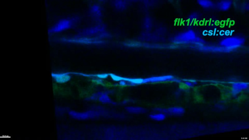
English: Movie 5 : Some flk1/kdrl: GFP;csl:Cer double-positive haemogenic endothelial cells leave the endothelium apically. This movie supports findings reported in Figure 1. It shows data from timelapse confocal microscopy on flk1/kdrl: gfp/csl: cer-double transgenic embryos starting from 30 hpf. The arrow points at a haemogenic endothelial cell that leaves the endothelium apically to enter the dorsal aorta. The embryo was immobilized in 1% low melting point agarose. The images show single 1.5 µm thick optical sagittal sections of an embryo with anterior to the left and dorsal up. During the timelapse, images were taken every 3 min. |
||
| Date | |||
| Source | Video file from Thambyrajah R, Ucanok D, Jalali M, Hough Y, Wilkinson R, McMahon K, Moore C, Gering M (2016). "A gene trap transposon eliminates haematopoietic expression of zebrafish Gfi1aa, but does not interfere with haematopoiesis". Developmental Biology. DOI:10.1016/j.ydbio.2016.07.010. PMID 27432513. PMC: 5003831. | ||
| Author | Thambyrajah R, Ucanok D, Jalali M, Hough Y, Wilkinson R, McMahon K, Moore C, Gering M | ||
| Permission (Reusing this file) |
This file is licensed under the Creative Commons Attribution 4.0 International license.
|
||
| Provenance |
|
File history
Click on a date/time to view the file as it appeared at that time.
| Date/Time | Thumbnail | Dimensions | User | Comment | |
|---|---|---|---|---|---|
| current | 20:01, 29 October 2016 | 11 s, 960 × 540 (973 KB) | Open Access Media Importer Bot (talk | contribs) | Automatically uploaded media file from Open Access source. Please report problems or suggestions here. |
You cannot overwrite this file.
File usage on Commons
There are no pages that use this file.