Category:SVG animal anatomy
Jump to navigation
Jump to search
Subcategories
This category has the following 9 subcategories, out of 9 total.
B
- SVG Bilaterian body plan (15 F)
- SVG bird anatomy (96 F)
C
- SVG crustacean anatomy (28 F)
F
- SVG fish anatomy (127 F)
G
- SVG Gastropoda anatomy (20 F)
I
M
R
- SVG reptiles anatomy (17 F)
W
- SVG worm anatomy (21 F)
Media in category "SVG animal anatomy"
The following 42 files are in this category, out of 42 total.
-
 Anatomy of Anodonta cygnea.svg 410 × 234; 116 KB
-
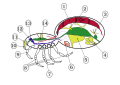 Araneae anatomic numbers.svg 744 × 524; 55 KB
-
 Araneae ru.svg 744 × 524; 66 KB
-
 Aranna pata.svg 562 × 422; 58 KB
-
 Archimollusc-fr.png 10,404 × 6,454; 11.7 MB
-
 Archimollusc-fr.svg 512 × 318; 1.91 MB
-
 Archimollusc.svg 2,492 × 1,536; 2.38 MB
-
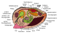
 Asterias rubens, dissection.svg 765 × 709; 674 KB
-
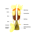 Bufonidae male reproductive system de.svg 1,000 × 1,000; 68 KB
-
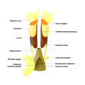 Bufonidae male reproductive system ru.svg 1,000 × 1,000; 69 KB
-
 Bumastus morphology.svg 874 × 543; 38 KB
-
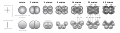
 Capas embrionarias.svg 843 × 690; 106 KB
-
 Cinctoblastula type of development Homoscleromorpha.svg 977 × 673; 612 KB
-
 Coeloblastula type of development Demospongiae.svg 978 × 305; 1.05 MB
-
 Collocyte - pl.svg 552 × 487; 65 KB
-
 Collocyte - ru.svg 552 × 487; 96 KB
-
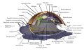
 Ctenophoran anatomy - ru.svg 795 × 1,125; 155 KB
-
 Ctenophore cleavage - ru.svg 1,500 × 410; 218 KB
-
 Ctenophore diagram - ru.svg 1,678 × 1,262; 162 KB
-
 Cuticula.svg 455 × 583; 61 KB
-
 Monoplacophora.svg 582 × 396; 18.91 MB
-
 Orthonectida anatomy - en.svg 670 × 636; 616 KB
-
 Orthonectida anatomy - es.svg 670 × 636; 700 KB
-
 Orthonectida anatomy - ru.svg 670 × 636; 616 KB
-
 Protovsdeuterostomes-de.svg 577 × 527; 1.09 MB
-
 Protovsdeuterostomes-es.svg 615 × 562; 1.16 MB
-
 Protovsdeuterostomes.svg 577 × 527; 1.31 MB
-
 Salticidae eye pattern.svg 330 × 237; 23 KB
-
 Schematic drawing of Amphiblastula type of development Calcarea.svg 1,048 × 592; 439 KB
-
 Schematic drawing of Calciblastula type of development Calcarea Calcinea.svg 1,056 × 697; 373 KB
-
 Schematic drawing of direct development Demospongiae Spirophorida.svg 1,034 × 717; 362 KB
-
 Schematic drawing of Disphaerula type of development Demospongiae.svg 1,000 × 676; 278 KB
-
 Schematic drawing of the first subtype of Parenchymella type of development.svg 1,058 × 818; 428 KB
-
 Schematic drawing of Trichimella type of development Hexactinellida - ru.svg 1,000 × 941; 448 KB
-
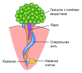
 Sea Anemone Structure.svg 512 × 384; 49 KB
-
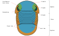
 Trilobite cephalon areas numbered.svg 260 × 180; 37 KB
-
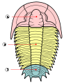
 Trilobite cephalon areas-en.svg 330 × 180; 31 KB
-
 Trilobite cranidium numbered.svg 230 × 160; 16 KB
-
 Trilobite cranidium-en.svg 270 × 190; 15 KB
-
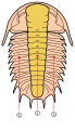 Trilobite lobes numbered.svg 217 × 376; 29 KB
-
 Trilobite sections numbered.svg 250 × 325; 38 KB
-
 卵割の様式.svg 1,600 × 1,100; 416 KB


