Category:On Molecular and Microscopic Science, Volume 1
Jump to navigation
Jump to search
Media in category "On Molecular and Microscopic Science, Volume 1"
The following 89 files are in this category, out of 89 total.
-
 I 004 - Eucyrtidium cranoides.jpg 323 × 500; 29 KB
-
 I 093 - Form of stratified discharge in a vacuum tube.jpg 300 × 84; 9 KB
-
 I 094.png 36 × 29; 2 KB
-

-
 I 185 - Development of Ulva.jpg 293 × 500; 39 KB
-
 I 187 - Vertical section of the cuticle of Iris germanica.jpg 300 × 162; 18 KB
-
 I 190 - Longitudinal section of stem of Italian reed.jpg 300 × 360; 33 KB
-
 I 196 - Palmoglœa macrococca.jpg 300 × 231; 18 KB
-
 I 198 - Protococcus pluvialis.jpg 300 × 304; 24 KB
-
 I 203 - Volvox globator.jpg 200 × 204; 17 KB
-
 I 206 - Various species of Staurastrum.jpg 400 × 286; 23 KB
-
 I 207 - Economy of Closterium Lunula.jpg 300 × 163; 12 KB
-
 I 211 - Diatoma vulgare and Grammatophora serpentina.jpg 343 × 400; 29 KB
-
 I 212 - Biddulphia pulchella.jpg 150 × 260; 7 KB
-
 I 213 - Pleurosigma angulatum.jpg 243 × 500; 25 KB
-
 I 214 - Actinocyclus undulatus.jpg 300 × 191; 19 KB
-
 I 215 - Meridion circulare.jpg 250 × 236; 20 KB
-
 I 217 - Bacillaria paradoxa.jpg 400 × 94; 9 KB
-
 I 221 - Cell multiplication in Conferva glomerata.jpg 248 × 400; 25 KB
-
 I 222 - Zoospores.jpg 250 × 275; 10 KB
-
 I 229 - Threads of Rivularia nitida.jpg 276 × 400; 22 KB
-
 I 230 - Trichodesmium erythræum.jpg 261 × 400; 21 KB
-
 I 231 - Conjugation of Zygnema quininum.jpg 400 × 167; 20 KB
-
 I 239 - Ulva latissima.jpg 400 × 308; 27 KB
-
 I 243 - Polyides rotundus and Furcellaria fastigiata.jpg 350 × 298; 28 KB
-
-
 I 246 - Callithamnion corymbosum.jpg 350 × 287; 21 KB
-

-
 I 256 - Dictyurus purpurascens.jpg 232 × 300; 18 KB
-
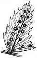
 I 257 - Polyzonia cuneifolia.jpg 400 × 320; 33 KB
-
 I 259 - Fruit of various species of Ectocarpus.jpg 400 × 282; 27 KB
-
 I 261 - Dictyota dichotoma.jpg 300 × 258; 19 KB
-
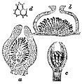
 I 268 - Vertical section of receptacle of Fucus platycarpus.jpg 338 × 500; 44 KB
-
 I 290 - Various species of Pucciniæi.jpg 332 × 400; 28 KB
-
 I 294 - Puccinia Graminis.jpg 198 × 300; 14 KB
-
 I 300 - Various species of Mucedines.jpg 400 × 222; 23 KB
-

-
 I 308 - Various species of Sphæriacei.jpg 300 × 318; 20 KB
-
 I 313 - Various species of Lichens.jpg 400 × 347; 26 KB
-

-

-
 I 326 - Nitella flexilis.jpg 173 × 500; 14 KB
-
 I 328 - Antheridia of Chara fragilis.jpg 400 × 223; 22 KB
-
 I 329 - Further development of antheridia of Chara fragilis.jpg 300 × 258; 18 KB
-
 I 331 - Marchantia polymorpha.jpg 300 × 226; 19 KB
-
 I 332 - Anatomy of frond of Marchantia polymorpha.jpg 500 × 168; 27 KB
-
 I 333 - Archegonia of Marchantia polymorpha.jpg 300 × 272; 18 KB
-
 I 333-1 - Elater and spores of Marchantia.jpg 80 × 600; 11 KB
-
 I 338 - Funaria hygrometrica.jpg 232 × 300; 13 KB
-
 I 339 - Polytrichum commune, group of antheridia.jpg 300 × 338; 23 KB
-
 I 339-1 - Polytrichum commune, development of spermatozoids.jpg 300 × 181; 14 KB
-
 I 344 - Microscopic structure of leaves of mosses.jpg 400 × 248; 23 KB
-
 I 351 - Development of spores of Pteris serrulata.jpg 400 × 316; 30 KB
-
 I 352 - Antheridium and spermatozoids of Pteris serrulata.jpg 300 × 148; 11 KB
-
 I 352-1 - Archegonium of Pteris serrulata.jpg 300 × 164; 12 KB
-
 I 355 - Section of footstalk of fern frond.jpg 250 × 280; 21 KB
-
 I 356 - Pinnule of Polypodium bearing sori.jpg 142 × 350; 14 KB
-
 I 357 - Sporangia of Polypodiaceous ferns.jpg 250 × 282; 22 KB
-
 I 360 - Pinnule of Lastrea Filix-mas with sori.jpg 200 × 286; 14 KB
-
 I 361 - Sorus and indusium of Polystichum or Aspidium.jpg 200 × 181; 13 KB
-
 I 362 - Pinna of Polystichum Lonchitis.jpg 194 × 300; 18 KB
-
 I 364 - Sorus and cup-shaped indusium of Deparia prolifera.jpg 250 × 237; 16 KB
-
 I 365 - Scolopendrium vulgare.jpg 250 × 153; 13 KB
-
 I 367 - Athyrium Filix-fœmina.jpg 200 × 221; 12 KB
-
 I 367-1 - Asplenium Ruta-muraria.jpg 200 × 227; 14 KB
-
 I 369 - Ceterach officinarum.jpg 200 × 259; 16 KB
-
 I 371 - Blechnum Spicant.jpg 174 × 250; 11 KB
-
 I 371-1 - Pteris aquilina.jpg 200 × 225; 12 KB
-
 I 373 - Adiantum Capillus-Veneris.jpg 300 × 165; 15 KB
-
 I 375 - Trichomanes radicans.jpg 125 × 200; 8 KB
-
 I 376 - Hymenophyllum tunbridgense.jpg 250 × 273; 16 KB
-
 I 382 - Equisetum giganteum.jpg 300 × 266; 18 KB
-
 I 385 - Pilularia minuta.jpg 86 × 200; 6 KB
-
 I 403 - Orchis mascula, side view of flower.jpg 400 × 295; 18 KB
-
 I 404 - Orchis mascula, front view of flower.jpg 256 × 300; 19 KB
-
 I 405 - Orchis mascula, pollinia.jpg 200 × 130; 8 KB
-
 I 405-1 - Orchis mascula, pollinium.jpg 84 × 200; 5 KB
-
 I 406 - Orchis mascula, pollen grains.jpg 200 × 215; 11 KB
-
 I 407 - Orchis pyramidalis, front view of flower.jpg 231 × 300; 15 KB
-
 I 408 - Orchis pyramidalis, side view of flower.jpg 339 × 400; 19 KB
-
 I 409-1 - Orchis pyramidalis, pollinia, withdrawn.jpg 200 × 217; 8 KB
-
 I 409a - Orchis pyramidalis, disc with one pollinium.jpg 150 × 214; 8 KB
-
 I 409b - Orchis pyramidalis, pollinia, attached to disc.jpg 146 × 200; 7 KB
-

-
 I 411 - Epipactis palustris, side views of flower.jpg 300 × 317; 16 KB
-
 I 412 - Epipactis palustris, side view and dissection of flower.jpg 400 × 250; 17 KB
-
 I 413 - Listera ovata, side view of flower.jpg 225 × 500; 25 KB
-
 Molecular And Microscopic Science Vol.1 (IA in.ernet.dli.2015.42261).pdf 695 × 1,056, 454 pages; 32.88 MB
-
 On Molecular and Microscopic Science, Volume 1.jpg 539 × 800; 47 KB


