Category:Microscopic images of leaves - trichomes
Jump to navigation
Jump to search
Subcategories
This category has the following 6 subcategories, out of 6 total.
M
S
Media in category "Microscopic images of leaves - trichomes"
The following 85 files are in this category, out of 85 total.
-
 Alchemilla SEM Stereo 200x.JPG 2,556 × 2,008; 2.44 MB
-
 Alchemilla SEM Stereo 500x.JPG 2,503 × 1,990; 2.15 MB
-
 Alchemilla SEM Stereo 50x.JPG 2,486 × 1,998; 1.89 MB
-
 Ampelocissus asekii trichomes - PhytoKeys-021-001-g003-C.jpeg 651 × 507; 163 KB
-
 Ampelocissus asekii trichomes - PhytoKeys-021-001-g003-E.jpeg 656 × 505; 147 KB
-
 Angiosperm Leaf Secondary Vascular Bundles in Nerium (24010128908).jpg 3,264 × 1,840; 4.74 MB
-
 Angiosperm Leaf Stomatal Pits in Nerium (24010130808).jpg 3,264 × 1,840; 4.6 MB
-
 Angiosperm Morphology Epidermal Trichome in Zea Leaf (37215761090).jpg 3,264 × 1,840; 938 KB
-

-
 Angiosperm Morphology Peltate Trichome in Syringa Epidermis (36840977652).jpg 3,264 × 1,840; 4.53 MB
-
 Archives de l'Institut botanique de l'Universit de Lige" (1897-) (19566111039).jpg 1,748 × 2,616; 847 KB
-
 Artemisia absinthium sl13.jpg 2,998 × 3,636; 2.4 MB
-
 Artemisia absinthium sl14.jpg 6,508 × 3,328; 5.73 MB
-
 Artemisia absinthium sl15.jpg 6,576 × 3,324; 5.73 MB
-
 Artemisia absinthium sl17.jpg 3,452 × 3,432; 3.29 MB
-
 Berteroa incana sl3.jpg 3,000 × 3,036; 2.89 MB
-
 Berteroa incana sl4.jpg 2,986 × 3,108; 3.25 MB
-
 Cannabis glandular trichomes.jpg 1,024 × 768; 167 KB
-
 Chenopodium vulvaria sl125.jpg 3,047 × 3,454; 4.43 MB
-
 Chenopodium vulvaria sl126.jpg 3,053 × 1,792; 445 KB
-
 Chenopodium vulvaria sl127.jpg 2,926 × 2,220; 532 KB
-
 Chenopodium vulvaria sl128.jpg 2,833 × 2,360; 633 KB
-
 Chenopodium vulvaria sl129.jpg 2,728 × 2,544; 595 KB
-
 Chenopodium vulvaria sl130.jpg 3,046 × 2,134; 598 KB
-
 Clover Leaf 60x.jpg 640 × 480; 133 KB
-
 Coleus Leaf Trichomes (hairs on the leaves) via Electron Microscope.jpg 1,024 × 1,049; 204 KB
-
 Coleus leaf trichomes SEM.jpg 1,024 × 1,049; 315 KB
-
 Crop of Elaeagnus umbellata leaf upper surface detail.jpg 367 × 496; 239 KB
-
 Detail, Walnut leaf 23 001 (cropped).jpg 2,670 × 3,335; 4.71 MB
-
 Dionaea-muscipula-Ausloeseborste-Mikroskopaufnahme.jpg 758 × 561; 111 KB
-
 Hippophae rhamnoides, Sanddorn,Trichom, pol. Licht.jpg 2,000 × 1,294; 1.58 MB
-
 Hydrillaverticillataleaf400x6.jpg 1,024 × 768; 57 KB
-
 Jatropha gossypiifolia - glandular petiole (4537141022).jpg 2,293 × 930; 417 KB
-
 Lavender leaf.jpg 600 × 425; 68 KB
-
 Leaf 123.jpg 1,176 × 999; 458 KB
-
 Leaf epidermis w scale.jpg 2,048 × 2,052; 1.1 MB
-
 Leaf epidermis.jpg 2,048 × 2,073; 2.9 MB
-
 Leaf Epidermis.jpg 720 × 960; 84 KB
-
 Levkoje o.jpg 404 × 481; 85 KB
-
 Levkoje sn.jpg 692 × 490; 221 KB
-
 Lobularia maritima sl1.jpg 3,014 × 2,871; 2.59 MB
-
 Microscopy image of a leaf.JPG 2,144 × 2,775; 555 KB
-
 Natural fence.jpg 3,456 × 2,000; 1.91 MB
-
 Nerium eingesenkte -Spaltoe.jpg 600 × 399; 169 KB
-
 Olive leaf lower surface 100x - SEM MUSE.tif 2,048 × 1,536; 3 MB
-
 Olive leaf lower surface 469x - SEM MUSE.tif 2,048 × 1,536; 3 MB
-
 Pelarg5.JPG 2,048 × 1,536; 774 KB
-
 Primula auricula sl11.jpg 3,096 × 3,053; 1.41 MB
-
 Primula auricula sl12.jpg 3,076 × 3,060; 2.08 MB
-
 Primula auricula sl13.jpg 3,074 × 3,091; 1.69 MB
-
 Primula auricula sl14.jpg 3,068 × 3,102; 1.55 MB
-
 Rosemary leaf lower surface 500x - SEM MUSE.tif 2,048 × 1,536; 3 MB
-
 Rumex sp. 10.JPG 1,791 × 1,340; 1.5 MB
-
 Rumex sp. 7.JPG 2,672 × 1,776; 556 KB
-
 Saxifraga tridactylites sl33.jpg 2,954 × 3,493; 1.52 MB
-
 Saxifraga tridactylites sl34.jpg 3,064 × 3,669; 1.07 MB
-
 Saxifraga tridactylites sl35.jpg 3,516 × 3,482; 1.38 MB
-
 Saxifraga tridactylites sl36.jpg 3,428 × 3,427; 1.29 MB
-
 Saxifraga tridactylites sl37.jpg 3,028 × 3,377; 1.24 MB
-
 Schmalblättrige Ölweide - Blattoberseite mit Sternhaaren.png 1,600 × 1,200; 3.25 MB
-
 Schmalblättrige Ölweide - Blattunterseite mit Sternhaaren.png 1,600 × 1,200; 3.17 MB
-
 Sesamothamnus lugardii 01.jpg 3,072 × 2,048; 6.41 MB
-
 Sesamothamnus lugardii 03.jpg 3,072 × 2,048; 5.63 MB
-
 Sesamum senecioides ab-01.jpg 4,992 × 3,328; 12.08 MB
-
 Sesamum senecioides ab-05.jpg 4,992 × 3,328; 11.88 MB
-
 Surface feuille de menthe poivree glandes secretrices.jpg 1,016 × 694; 85 KB
-
 Tobacco leaf surface micro photo.jpg 3,840 × 2,880; 2.55 MB
-
 Tricoma digitiforme (folha de Mentha) (2969298877).jpg 800 × 600; 69 KB
-
 Urtica dioica hairs.jpg 1,280 × 960; 566 KB
-
 Veronica sublobata sl28.jpg 3,372 × 3,410; 2.75 MB
-
 Veronica sublobata sl29.jpg 3,384 × 3,449; 2.34 MB
-
 Walnut leaf 23 001.jpg 4,120 × 4,095; 8.89 MB
-
 Walnut leaf 23 002.jpg 4,100 × 4,125; 8.79 MB
-
 Wet bottle brush leaf stomata and leaf hairs no scale bar.jpg 3,385 × 2,462; 3.41 MB
-
 Wet bottle brush leaf stomata and leaf hairs.jpg 600 × 436; 63 KB
-
 Волоски листа герани под микроскопом.jpg 833 × 1,058; 33 KB
-
 Волоски на поверхности листа.tif 1,024 × 943; 2.77 MB
-
 Різновиди трихом-емергенець.jpg 2,005 × 1,944; 2.16 MB
-
 Секреторные волоски на листе герани. Автофлуоресценция 2.tif 2,780 × 2,780; 44.24 MB
-
 Секреторные волоски на листе герани. Автофлуоресценция 3.tif 3,472 × 3,472; 69 MB
-
 Секреторные волоски на листе герани. Автофлуоресценция 4.tif 4,708 × 3,139; 84.58 MB
-
 Секреторные волоски на листе герани. Автофлуоресценция 5.tif 4,482 × 2,988; 76.65 MB
-
 Секреторные волоски на листе герани. Автофлуоресценция.tif 3,232 × 3,232; 59.79 MB
-
 Трихома на листе лимонной герани.tif 5,173 × 3,448; 51.05 MB
-
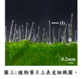 植物葉片上表皮組織圖.png 559 × 537; 486 KB


.jpg)
.jpg)
.jpg)
.jpg)
.jpg)
_(19566111039).jpg)


_via_Electron_Microscope.jpg)


.jpg)


_(2969298877).jpg)


.jpg)