Category:Images from Scientific Images album of National Center for Advancing Translational Sciences Flickr stream uploaded by Netha Hussain
Jump to navigation
Jump to search
Media in category "Images from Scientific Images album of National Center for Advancing Translational Sciences Flickr stream uploaded by Netha Hussain"
The following 54 files are in this category, out of 54 total.
-
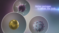 23 Better Packages to Deliver the Tools (48908962132).png 1,920 × 1,080; 1.35 MB
-
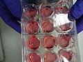
 3-D Bioprinted Tissues in Well Plate (42569479372).jpg 659 × 436; 108 KB
-
 3-D Bioprinting (42479582471).jpg 550 × 337; 223 KB
-
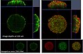
 3-D Image of Bioprinted Skin Tissue (28746740398).jpg 1,289 × 725; 133 KB
-
 3-D Image of Cancer Tumor (40813889460).jpg 930 × 610; 94 KB
-
 3-D Printed Eye Tissue (34100509955).jpg 4,864 × 3,648; 3.93 MB
-
 3D system human gut.png 556 × 411; 360 KB
-
 AAV micro-dystrophin gene therapy (36049415430).jpg 382 × 626; 54 KB
-
 An Itch to Scratch (48905779046).jpg 483 × 483; 294 KB
-
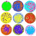 Analyzing Immune Activation (33597341278).jpg 2,883 × 2,919; 840 KB
-
 Barth syndrome.png 556 × 402; 129 KB
-
 Bioengineered blood vessel.png 556 × 292; 184 KB
-
 Bioprinting Cellular Patterns (40812191430).jpg 211 × 212; 21 KB
-
 Brain Cells (40522751690).png 556 × 615; 653 KB
-
 Brain Chip (40529717123).jpg 591 × 592; 48 KB
-
 Brain Chip (40529718453).jpg 960 × 882; 265 KB
-
 Brain Chip (40538388250).jpg 556 × 541; 109 KB
-
 Brain Chip (46771920474).jpg 674 × 526; 104 KB
-
 Brain Tissue Cells (33272648413).png 556 × 310; 326 KB
-
 Brain Tumor on Brain Slice (27588729787).jpg 1,152 × 1,152; 774 KB
-
 Cells in Simulated Microgravity (33597341708).png 692 × 583; 452 KB
-
 Computed 3-D Tumor Sphere (42569469392).jpg 674 × 626; 138 KB
-
 Cyclic peptide ipglycermide.png 1,250 × 1,017; 912 KB
-
 DNA Repair Enzyme Binds to DSB (48908756201).png 1,920 × 1,080; 2.43 MB
-
 Duchenne Muscular Dystrophy (35122690776).jpg 1,024 × 1,024; 196 KB
-
 Embryoid Body Formation (33597343448).jpg 1,600 × 1,200; 956 KB
-
 Fluorescent green lipid.png 556 × 140; 82 KB
-
 Gene therapy approach for Pompe disease(28389581788).jpg 900 × 600; 123 KB
-
 Heart Chip (33970475351).png 556 × 172; 278 KB
-
 Histology of 3-D Bioprinted Skin (42621985651).jpg 9,576 × 3,102; 1.97 MB
-
 Human Cardiac Tissue Chip (47473708501).jpg 1,022 × 935; 296 KB
-
 Human colon tumor.png 556 × 521; 459 KB
-
 Human Eosinophils White Blood Cells (42037940875).jpg 900 × 600; 190 KB
-
 Human Gut Tissue Chip (46750334684).png 1,030 × 556; 869 KB
-
 Human Lung Airway Chip (46558399795).png 322 × 432; 359 KB
-
 Human Muscle Chip (47420765402).jpg 1,024 × 1,024; 262 KB
-
 Human Stem Cells (40529583203).jpg 785 × 1,029; 837 KB
-
 Hunter Syndrome Brain (41772505464).jpg 900 × 600; 89 KB
-
 Huntington’s disease (40456396870).jpg 900 × 600; 65 KB
-
 Integrated Multi Chip (36896123285).jpg 4,620 × 3,646; 2.8 MB
-
 Kidney Mini-Organs Zoom (46580295955).jpg 1,600 × 808; 697 KB
-
 Kidney Organoids (46580296595).jpg 1,600 × 1,105; 713 KB
-
 Kidney-on-a-Chip (33595959148).jpg 3,319 × 2,489; 4.85 MB
-
 Kidney-on-a-Chip (47419676542).jpg 5,486 × 3,657; 15.17 MB
-
 Liver Cancer in Mouse Model (42507312762).jpg 786 × 715; 113 KB
-
 Liver Chip (33258222594).png 556 × 277; 216 KB
-
 Lymphangioleiomyomatosis .png 424 × 281; 336 KB
-
 Microguts (33258222124).png 556 × 211; 119 KB
-
 Microvessel networks.png 556 × 174; 147 KB
-
 Mini-Brains (42329302011).jpg 450 × 449; 203 KB
-
 Mouse Brain (48908239833).jpg 2,056 × 2,056; 1.39 MB
-
 Ovarian Cancer Cells (34043700576).jpg 301 × 313; 14 KB
-
 Sickle cells illustration.jpg 578 × 439; 55 KB
-
 Small Molecule Probe Targeting Cancer (41658265005).jpg 1,024 × 1,024; 69 KB
.png)
.jpg)
.jpg)
.jpg)
.jpg)
.jpg)

.jpg)
.jpg)
.jpg)


.jpg)
.png)
.jpg)
.jpg)
.jpg)
.jpg)
.png)
.jpg)
.png)
.jpg)

.png)
.jpg)
.jpg)
.jpg)
.jpg)

.jpg)
.png)
.png)
.jpg)
.jpg)
.jpg)
.jpg)
.jpg)
.jpg)
.jpg)
.jpg)
.jpg)
.jpg)
.png)

.jpg)
.jpg)
.jpg)

.jpg)
.png)
.jpg)
.png)