Category:Human anatomical models
Jump to navigation
Jump to search
Subcategories
This category has the following 15 subcategories, out of 15 total.
Media in category "Human anatomical models"
The following 136 files are in this category, out of 136 total.
-
 3-D Printed Skull Plate (5255) (18492487005).jpg 3,076 × 2,311; 3.9 MB
-
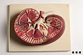
 4167134294 d0cf029385 bTêteCoupe.jpg 2,451 × 3,050; 487 KB
-
 A human hand anatomised and preserved Wellcome L0036404.jpg 3,008 × 1,960; 727 KB
-
 A medical student before exam in saloon of moulages.JPG 1,472 × 2,392; 898 KB
-
 Adonis in Wachs.JPG 960 × 1,280; 285 KB
-
 AED & ICD Research (5334) (18304620558).jpg 1,842 × 1,330; 1.63 MB
-
 Anatomical ivory model of ear Wellcome L0010001.jpg 1,558 × 1,268; 294 KB
-
 Anatomical model of human brain, Wellcome L0010008.jpg 1,474 × 1,186; 469 KB
-
 Anatomical model of human brain, Wellcome L0010009.jpg 1,440 × 1,216; 467 KB
-
 Anatomical model. Wellcome L0012201.jpg 882 × 1,995; 705 KB
-
 Anatomical Venus. Wax figure of reclining woman, Florence. Wellcome L0058207.jpg 4,256 × 2,832; 821 KB
-
 Anatomy (6941784778).jpg 3,209 × 4,844; 3.49 MB
-
 Anatomy model LPB Laos.jpg 1,536 × 1,024; 391 KB
-
 Auditory System Animation.gif 1,454 × 799; 385 KB
-
 Auzoux papiermache model van zeebaars 20701.JPG 1,004 × 1,500; 464 KB
-
 Auzoux zwangerschapsmodel papiermache 31932-b.JPG 1,500 × 1,000; 399 KB
-
 Back Anatomy and Medulla Spinalis.jpg 3,587 × 5,259; 3.07 MB
-
 Back Anatomy and Medulla Spinalis2.jpg 3,653 × 5,479; 3.13 MB
-
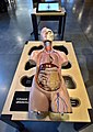
 BLW Human Anatomy.jpg 2,304 × 3,456; 3.56 MB
-
 Brüste im Modell - breasts in the model.jpg 3,264 × 2,448; 1.61 MB
-
-

-
 Cap anatòmic, c. 1850 - 1860, col·lecció cientificomèdica, Universitat de València.JPG 2,793 × 3,867; 1.27 MB
-

-
 Centrum Nauki Kopernik człowiek układanka 2019g.jpg 4,875 × 6,899; 3.31 MB
-
 Chest Anatomy.jpg 5,616 × 3,744; 3.88 MB
-
 Chiropractor - DPLA - 92d3e67cd3496fa68b28390a5a2d8de4.jpg 2,848 × 4,256; 4.19 MB
-
 Człowiek-układanka. Obiekt na wystawie w Centrum Nauki Kopernik w Warszawie.jpg 2,818 × 3,134; 5.25 MB
-
 Darmmodell.JPG 570 × 858; 87 KB
-
 Didactic model of a human embryonic development 01.jpg 3,872 × 2,592; 1.48 MB
-
 Didactic model of a human embryonic development 02.jpg 3,767 × 2,522; 2.48 MB
-
 Didactic model of a human embryonic development 03.jpg 3,791 × 2,538; 2.94 MB
-
 Didactic model of a human embryonic development 04.jpg 3,872 × 2,592; 2.9 MB
-
 Didactic model of a human embryonic development 05.jpg 3,872 × 2,592; 2.79 MB
-
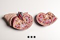 Didactic model of a mammal Kidney and adrenal gland.jpg 3,872 × 2,592; 4.41 MB
-
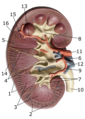 Didactic model of a mammal Kidney with numbered parts.png 2,592 × 3,644; 10.03 MB
-
 Didactic model of a mammal kidney-FMVZ USP-16.jpeg 3,763 × 2,519; 4.73 MB
-
 Didactic model of a mammal Kidney.jpg 3,872 × 2,592; 5.34 MB
-
 Didactic model of respiratory system-FMVZ USP-12.jpeg 3,872 × 2,592; 4.15 MB
-
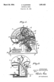 Dr. Edith Klemperer Patent for Luminous Brain Model, 1934.png 407 × 615; 116 KB
-
 Basel 2012-10-05 Batch 2 (21).JPG 2,736 × 3,648; 3.06 MB
-
 Basel 2012-10-05 Batch 2 (22).JPG 2,736 × 3,648; 3.12 MB
-
 Basel 2012-10-05 Batch 2 (23).JPG 3,648 × 2,736; 3.61 MB
-
 Ecorchefragonard20100711.JPG 4,000 × 3,000; 2.6 MB
-
 Eternal breath.JPG 5,184 × 3,456; 3.92 MB
-
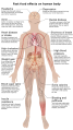 Fast food effects on human body (en).svg 1,363 × 2,420; 2.36 MB
-
 Fast food effects on human body (it).svg 1,363 × 2,420; 2.51 MB
-
 Follow me, the exit is up there ! (38484588644).jpg 3,300 × 4,938; 3.48 MB
-

-
 Gehirnmodell.JPG 600 × 488; 112 KB
-
 Human anatomy planes, labeled.jpg 4,280 × 3,696; 914 KB
-
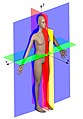 Human anatomy planes.jpg 2,524 × 3,624; 495 KB
-
 Human body by rua.jpg 720 × 1,280; 156 KB
-
 Human brain 01.jpg 2,338 × 3,492; 3.67 MB
-
 Human brain 02.jpg 2,109 × 3,150; 2.93 MB
-
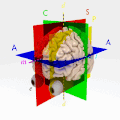
 Human brain anatomical planes letter annotations.jpg 1,751 × 1,583; 283 KB
-
 Human ear (teaching model showing internal mechanisms).jpg 3,872 × 2,592; 4.37 MB
-
 Human eyeball.jpg 2,368 × 3,537; 3.36 MB
-
 Human facial vascularization.jpg 2,368 × 3,537; 3.22 MB
-
 Human head anatomical planes letter annotations.jpg 2,592 × 2,482; 542 KB
-
 Human head with labeled anatomic planes.jpg 4,000 × 4,000; 384 KB
-
 HumanModelSilikon.jpg 4,000 × 6,000; 18.12 MB
-
 Il corpo umano.jpg 6,000 × 4,000; 4.39 MB
-
 Il cuore.jpg 4,608 × 3,072; 2.91 MB
-
 Inauguración Centro de Estudios Navales en Ciencias de la Salud (30378328808).jpg 1,994 × 1,494; 1.5 MB
-
 Inauguración Centro de Estudios Navales en Ciencias de la Salud. (43339565295).jpg 1,935 × 1,354; 1.66 MB
-
 Ivory and horn model of an eye, Europe, 1801-1900 Wellcome L0057567.jpg 4,256 × 2,832; 810 KB
-
 Ivory model of a human ear, Europe, 1701-1800 Wellcome L0057746.jpg 3,430 × 4,529; 1.48 MB
-
 Le musée danatomie humaine (Turin) (2863076603).jpg 861 × 1,200; 482 KB
-
 Male anatomical figure in wood Wellcome M0018671EA.jpg 1,092 × 3,733; 1.1 MB
-
 Male anatomical figure in wood Wellcome M0018671EB.jpg 2,197 × 4,845; 2.19 MB
-
 Materials of the human body.jpg 3,434 × 2,984; 1.02 MB
-
 Medical students before exam in saloon of moulages 1.JPG 2,816 × 2,112; 2.79 MB
-
 Medical students before exam in saloon of moulages2.JPG 1,312 × 796; 337 KB
-

-
 Model of a human brain, Europe, 1801-1850 Wellcome L0057095.jpg 4,256 × 2,832; 1.26 MB
-
 Model of a human brain, France, 1801-1850 Wellcome L0057743.jpg 3,528 × 4,704; 2.36 MB
-
 Model of an eye, Europe, 1801-1900 Wellcome L0058736.jpg 2,832 × 4,012; 1.18 MB
-
 Model.gif 236 × 167; 23 KB
-
 Modell eines menschlichen Ohrs.jpg 3,072 × 2,304; 3.62 MB
-

-
 Moscow, VDNKh, anatomical models display (10656984353).jpg 5,472 × 3,648; 2.15 MB
-
 Moulage of ear.JPG 1,536 × 2,576; 869 KB
-
 Moulage of muscles of face.JPG 1,504 × 1,848; 787 KB
-
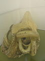
 Moulage of skull base.JPG 1,984 × 2,512; 1.15 MB
-
 Musée de l'Homme Cire anatomique Artères et nerfs Carlo Calenzuoli 04022018 1.jpg 4,590 × 3,151; 5.13 MB
-
 Musée de l'Homme Cire anatomique Hémi-crâne 04022018 1.jpg 2,323 × 3,458; 3.3 MB
-
 Musée de l'Homme Cire Myologie face et cou Carlo Calenzuoli 04022018 2.jpg 3,728 × 2,909; 4.42 MB
-
 Musée de l'Homme Cire Myologie face et cou Carlo Calenzuoli 04022018 3.jpg 4,752 × 3,168; 5.61 MB
-
 Musée de l'Homme Cire Myologie face et cou Carlo Calenzuoli 04022018.jpg 3,685 × 2,242; 3.68 MB
-
 Obstetrical room - Palazzo Poggi.jpg 3,482 × 2,591; 1.77 MB
-
 ParasagittalBrain.jpg 4,000 × 4,000; 587 KB
-
 PlanesOfSection.jpg 2,248 × 2,349; 250 KB
-
 Probing-procedure.jpg 708 × 531; 115 KB
-
-

-

-

-

-

-

-
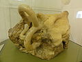
 Resin skull (3081230096).jpg 2,642 × 1,925; 1.51 MB
-
 Respiratory function by Bryan Brandenburg.jpg 1,200 × 1,200; 193 KB
-
 RotatingParasagittalBrain.gif 320 × 320; 1.32 MB
-
 Skelett im Anatomischen Museum Basel - 4675.jpg 638 × 1,798; 1.23 MB
-
 Skinless head sideview.jpg 1,071 × 1,428; 335 KB
-
 Specola 1.jpg 1,539 × 1,200; 449 KB
-
 Specola 10.jpg 1,236 × 1,200; 388 KB
-
 Specola 11.jpg 1,116 × 1,470; 394 KB
-
 Specola 12.jpg 1,200 × 1,600; 524 KB
-
 Specola 13.jpg 1,200 × 1,600; 468 KB
-
 Specola 14.jpg 1,113 × 1,509; 424 KB
-
 Specola 16.jpg 1,068 × 1,527; 439 KB
-
 Specola 17.jpg 1,122 × 1,569; 438 KB
-
 Specola 18.jpg 718 × 956; 187 KB
-
 Specola 19.jpg 1,068 × 1,032; 285 KB
-
 Specola 22.jpg 765 × 1,026; 202 KB
-
 Specola 24.jpg 1,598 × 1,118; 1.02 MB
-
 Specola 25.jpg 1,092 × 1,515; 460 KB
-
 Specola 26.jpg 1,125 × 1,554; 442 KB
-
 Specola 4.jpg 1,119 × 1,425; 432 KB
-
 Specola 6.jpg 1,600 × 1,200; 525 KB
-
 Specola 7.jpg 864 × 1,191; 269 KB
-
 Specola 8.jpg 1,032 × 1,284; 368 KB
-
 Specola 9.jpg 1,600 × 1,200; 505 KB
-
 Spinal readjustment 3.jpg 447 × 497; 34 KB
-
 The female Anatomy.jpg 3,024 × 4,032; 3.15 MB
-
 The male Anatomy.jpg 3,024 × 4,032; 2.96 MB
-
 Tonstudio 1985 Technische Sammlungen Dresden 02.JPG 4,000 × 2,248; 3.9 MB
-
 Urban legends regarding masturbation.png 658 × 493; 202 KB
-

-
 Wax injected human left arm, Europe, 1831-1870 Wellcome L0057342.jpg 2,832 × 4,256; 688 KB
-
 Whites’ Physiological manikin..JPG 695 × 768; 294 KB
-
 Wooden Anatomical Figures Wellcome L0027917.jpg 2,882 × 3,890; 2.99 MB
-
 Zoologia La Specola - wax anatomical models.JPG 2,560 × 1,920; 832 KB
-
 مجسم لجسم الإنسان.jpg 720 × 1,280; 75 KB
_(18492487005).jpg)


_(18304620558).jpg)


.jpg)


.jpg)


.JPG)
.JPG)
.JPG)


.svg)
.svg)
.jpg)
_by_Dr_John_Hunter,_Hunterian_Art_Gallery,_Glasgow.jpg)


.jpg)


.jpg)
.jpg)


_(2863076603).jpg)


.jpg)


.jpg)


.jpg)


