Category:Fluorescence microscopic images with mitochondria specific stains
Jump to navigation
Jump to search
Media in category "Fluorescence microscopic images with mitochondria specific stains"
The following 33 files are in this category, out of 33 total.
-
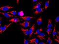
 AMA ANTIBODIES.jpg 1,877 × 1,533; 4.09 MB
-

-

-

-

-

-

-
 Bovine Pulmonary Artery Endothelial Cells Fluorescent Image 2.jpg 1,920 × 1,452; 298 KB
-
 Bovine Pulmonary Artery Endothelial Cells Fluorescent Image 3.jpg 1,920 × 1,452; 423 KB
-
 Bovine Pulmonary Artery Endothelial Cells Fluorescent Image.jpg 1,920 × 1,452; 304 KB
-
 C1orf112SubcellularLocalization.jpg 200 × 200; 13 KB
-
 DAPIMitoTrackerRedAlexaFluor488BPAE.jpg 1,384 × 1,040; 1.41 MB
-
 Fibroblast (BPAE).jpg 3,072 × 2,048; 2.79 MB
-
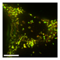
 Fibroblast,polydopamine nanoparticles and mithocondria.jpg 2,048 × 2,048; 579 KB
-
 Fibroblastid (BPAE).jpg 3,304 × 2,202; 2.73 MB
-
 Fibroblastid.jpg 6,000 × 4,627; 7.53 MB
-
 HeLa mtGFP.tif 1,024 × 1,024; 3 MB
-
 HeLa-Tubulin-HSP60-Fibrillarin-DNA.jpg 2,968 × 2,976; 4.93 MB
-
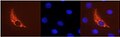
 HSP60-1C7.jpg 1,600 × 1,200; 223 KB
-
 Indian Muntjac fibroblast cells (24271618921).jpg 1,923 × 1,210; 684 KB
-
 MEF cell mitochondria 2.jpg 1,024 × 1,024; 102 KB
-
 MEF cell mitochondria.jpg 1,024 × 1,024; 103 KB
-
 Mitochondria & Nucleus overlay.jpg 604 × 184; 10 KB
-
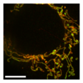
 Mitochondria in living HeLa cells.jpg 719 × 607; 27 KB
-
 Mitochondrial OMM and matrix in DRP1-KO cells after PXA.png 296 × 296; 88 KB
-
 Mitochondrial OMM and matrix in MEF DRP1-KO CCCP.png 295 × 296; 110 KB
-
 Mitochondrial OMM and matrix in MEF WT CCCP.png 296 × 296; 112 KB
-
 Mitochondrial OMM and matrix in MEF WT CTRL.png 296 × 296; 98 KB
-
 Mitokondrid, mikrofilamendid ja tuumad rakkudes.jpg 5,648 × 3,566; 9.9 MB
-
 Multicolor fluorescence image of a living HeLa cell.jpg 1,024 × 1,024; 148 KB
-
 Multicolor fluorescence image of a living PC-12 cell.jpeg 1,024 × 1,024; 79 KB
-
 Multicolor fluorescence image of living HeLa cells.jpg 1,024 × 1,024; 184 KB
-
 Snap-4064 21.tif 2,560 × 1,920; 4.16 MB


.jpg)

.jpg)


.jpg)


