User:Curtis Clark/gallery
Jump to navigation
Jump to search
Mon Jun 11 15:00:03 PDT 2012
[edit]-
Light micrograph of a whole-mount slide of an Antoceros gametophyte with embedded sporophyte
-
Light micrograph of a preserved specimen of an Antoceros gametophyte with embedded sporophytes
-
Light micrograph of a whole-mount slide of the base of an Anthoceros sporophyte embedded in the gametophyte
-
Light micrograph of a longitudinal section of Azolla, showing the cyanobacterium Anabaena in leaf pockets
-
Light micrograph of a cross section of the basidiocarp of Boletus, showing the hymenium lining the pores
-
Light micrograph of a cross section of the basidiocarp of Boletus, showing the pores
-
Light micrograph of a whole-mount slide of an oogonium and antheridium of Chara
-
Light micrograph of a cross section of the gills of Coprinus, showing the hymenium with basidia
-
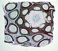
Light micrograph of a cross section of a coralloid root of a cycad, showing the layer that hosts symbiotic cyanobacteria
-
Light micrograph of a cross section of a coralloid root of a cycad, showing the layer that hosts symbiotic cyanobacteria
-
Light micrograph of a preserved specimen of coralloid roots of Cycas
-
Light micrograph of a cross section of the microsporophyll and microsporangia of Cycas
-
Light micrograph of a cross section of the microsporophyll and microsporangia of Cycas
-
Light micrograph of a whole-mount slide of the plurilocular structure of Ectocarpus
-
Light micrograph of a whole-mount slide of unilocular structures of Ectocarpus
-
Light micrograph of a whole-mount slide of a unilocular structure of Ectocarpus
-
Ephedra (probably E. californica) growing on the Carrizo Plain of California
-
Light micrograph of a longitudinal section of the ovule of Ephedra
-
Light micrograph of a whole-mount slide of gametangia of Equisetum
-
Light micrograph of a cross section of a conceptacle of Fucus, showing oogonia and antheridia
-
Light micrograph of a cross section of a conceptacle of Fucus, showing oogonia and antheridia
-
Light micrograph of a cross section of a conceptacle of Fucus, showing oogonia and antheridia
-
Light micrograph of a cross section of a receptacle of Fucus, showing conceptacles
-
Light micrograph of a cross section of a receptacle of Fucus, showing conceptacles
-
Light micrograph of a preserved specimen of the receptacle of Fucus, showing the openings of the conceptacles
-
Light micrograph of a preserved specimen of the receptacle of Fucus, showing the openings of the conceptacles
-
Light micrograph of a preserved specimen of the pollen cone of Ginkgo, showing microsporophylls with microsporangia
-
Light micrograph of a longitudinal section of the pollen cone of Ginkgo, showing microsporophylls with microsporangia and pollen
-
Light micrograph of a longitudinal section of the pollen cone of Ginkgo, showing microsporophylls with microsporangia and pollen
-
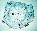
Light micrograph of a cross section of a young Ginkgo stem
-
Light micrograph of a longitudinal section of the blade of Laminaria, showing external meiosporangia and internal conductive tissue
-
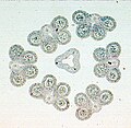
Light micrograph of a cross section of the anthers and style of a Lilium flower
-
Light micrograph of a cross section of an anther of a Lilium flower
-
Light micrograph of a preserved specimen of the fametophyte of Lycopodium, with embedded sporophyte
-
Light micrograph of a longitudinal section of a Lycopodium strobilus
-
Light micrograph of a longitudinal section of a Lycopodium strobilus
-
Light micrograph of a preserved specimen ofthe antheridiophores of Marchantia
-
Light micrograph of a preserved specimen of and archegoniophore of Marchantia, with embedded sporophytes
-
Living specimen of a germinated sporocarp of Marsilea, showing the axis and the megasporangia and microsporangia
-
Living specimen of a germinated sporocarp of Marsilea, showing the axis and the megasporangia and microsporangia
-
Living specimen of female gametophytes (inside megaspores) and male gametophytes (inside microspores) of Marsilea
-
Light micrograph of a cross section of a Marsilea sporocarp
-
Light micrograph of a whole-mount slide of a moss peristome
-
Light micrograph of a whole-mount slide of the oogonia and antheridia of Oedogonium
-
Light micrograph of a whole-mount slide of an oogonium and andtheridia of Oedogonium
-
Light micrograph of a cross section of an Osmunda stem
-
Pelagophycus porra washed up on the southwest coast of San Clemente Island, CA
-
Light micrograph of a cross section of a Phaseolus fruit
-
Light micrograph of a cross section of a Phaseolus fruit
-
Living specimen of Phycomyces zygospores
-
Living specimen of Phycomyces zygospores
-
Light micrograph of a cross section of the apothecium of Physcia
-
Light micrograph of a cross section of the apothecium of Physcia
-
Light micrograph of a longitudinal section of a pine female gametophyte and embryo
-
Light micrograph of a longitudinal section of a pine ovule, showing fertilization
-
Light micrograph of a longitudinal section of a pine ovule, showing fertilization
-
Light micrograph of a whole-mount slide of germinated pine pollen grains
-
Light micrograph of a longitudinal section of a pine pollen cone, showing microsporophylls and microsporangia
-
Light micrograph of a longitudinal section of a young pine seed cone, showing bracts, cone scales, and ovules
-
Light micrograph of a preserved specimen of a young pine seed cone
-
Light micrograph of a preserved specimen of a young pine seed cone, sectioned longitudinally
-
Light micrograph of a whole-mount slide of the cystocarp and carposporophyte of Polysiphonia
-
Light micrograph of a preserved specimen of cystocarps of Polysiphonia
-
Light micrograph of a whole-mount slide of spermatangia of Polysiphonia
-
Light micrograph of a whole-mount slide of spermatangia of Polysiphonia
-
Light micrograph of a whole-mount slide of tetraspores of Polysiphonia
-
Light micrograph of a whole-mount slide of tetraspores of Polysiphonia
-
Light micrograph of a preserved specimen of tetraspores of Polysiphonia
-
Light micrograph of a longitudinal section of the archegoniophore of Porella
-
Light micrograph of a whole-mount slide of the archegoniophore of Porella
-
Light micrograph of a preserved specimen of Porella with archegoniophores
-
Light micrograph of a preserved specimen of a Porella gametophyte with embedded sporophytes
-
Foliar appendage subtending the synangium of Psilotum nudum
-
Light micrograph of a preserved specimen of the rhizome of Psilotum nudum
-
Stem of Psilotum nudum with synangia
-
Light micrograph of a whole-mount slide of the clonal sporangia of Rhizopus
-
Light micrograph of a whole-mount slide of zygospores of Rhizopus
-
Light micrograph of a whole-mount slide of zygospores of Rhizopus
-
Light micrograph of a whole-mount slide of the endosporic female gametophyte of Selaginella with embedded embryonic sporophyte
-
Light micrograph of a longitudinal section of a sporophyll of Selaginella bearing a megasporangium with megaspores
-
Light micrograph of a longitudinal section of a Selaginella strobilus, showing microsporangia (left) and megasporangia (right)
-
Light micrograph of a longitudinal section of a Selaginella strobilus, showing microsporangia (right) and megasporangia (left)
-
Light micrograph of a whole-mount slide of a flower of Triticum
-
Light micrograph of a whole-mount slide of a flower of Triticum
-
Light micrograph of a longitudinal section of a caryopsis (grain) of Triticum
-
Light micrograph of a cross section of the thallus of Umbilicaria, showing a layer of algal cells in the upper portion
-
Light micrograph of a cross section of the thallus of Umbilicaria, showing a layer of algal cells in the upper portion
-
Light micrograph of a whole-mount slide of cleistothecia of Uncinula on a host plant leaf
-
Light micrograph of a longitudinal section of a Zamia ovule, shwoing eggs, female gametophyte, nucellus, and integument
-
Living specimen of a young fern gametophyte
-
Light micrograph of a whole-mount slide of
-
Light micrograph of a whole-mount slide of
-
Light micrograph of a tangential longitudinal section of Pinus wood
-
Light micrograph of a cross section of a young Pinus stem
-
Light micrograph of a cross section of a young Pinus stem
Sat Jul 05 15:40:27 PDT 2008
[edit]-
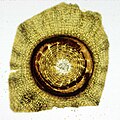
Diatom test in commercial diatomaceous earth for swimming pool filters
-
Diatom test in commercial diatomaceous earth for swimming pool filters
Sun May 04 20:35:49 PDT 2008
[edit]-
Eschscholzia caespitosa, Sierra Nevada foothills, California
-
”Eschscholzia hypecoides, California
-
Eschscholzia lemmonii, California
-
Eschscholzia minutiflora subsp. minutiflora, Mojave Desert, California
-
Eschscholzia minutiflora subsp. twisselmannii, Red Rock Canyon State Park, California
-
Eschscholzia ramosa, San Clemente Island, California
-
Eschscholzia rhombipetala", Carrizo Plain, California
Wed Jul 05 10:59:55 PDT 2006
-
Voorhis Ecological Reserve, near Pomona, California.
-
Voorhis Ecological Reserve, near Pomona, California.
-
Northeast of Valle, Arizona.
-
Northeast of Valle, Arizona.
-
Northeast of Valle, Arizona.
-
Northeast of Valle, Arizona.
-
Voorhis Ecological Reserve, near Pomona, California. Species is non-native.
-
Northeast of Valle, Arizona.
-
Grown in cultivation.
-
Top of Red Butte (Wii'i Gdwiisa), Coconino Co., Arizona.
-
Photographed at [BioTrek.
-
Voorhis Ecological Reserve, near Pomona, California.
-
Voorhis Ecological Reserve, near Pomona, California.
-
Northeast of Valle, Arizona. Species is a non-native weed.
-
Northeast of Valle, Arizona. Species is a non-native weed.
-
Northeast of Valle, Arizona.
Tue Jul 04 14:23:39 PDT 2006
-
Photographed in Voorhis Ecological Reserve near Pomona, CA, USA. The inflorescence is a cincinnus (scorpioid cyme).
-
Photographed in Voorhis Ecological Reserve near Pomona, CA, USA. Species is an introduced weed.
-
Photographed in Voorhis Ecological Reserve near Pomona, CA, USA.
-
Northeast of Valle, Arizona.
-
Photographed in Voorhis Ecological Reserve near Pomona, CA, USA. Species is an introduced weed.
-
Photographed in Voorhis Ecological Reserve near Pomona, CA, USA. Species is an introduced weed.
-
Photographed in Voorhis Ecological Reserve near Pomona, CA, USA.
-
Northeast of Valle, Arizona.
-
Northeast of Valle, Arizona.
-
Photographed in Voorhis Ecological Reserve near Pomona, CA, USA.
-
Photographed in Voorhis Ecological Reserve near Pomona, CA, USA.
-
Photographed at [BioTrek, Pomona, CA.
-
Photographed at [BioTrek, Pomona, CA.
-
East of Needles, CA. Species is an introduced weed.
-
San Gabriel Mts. north of Rancho Cucamonga, CA
-
Photographed in Voorhis Ecological Reserve near Pomona, CA, USA.
-
San Gabriel Mts. north of Rancho Cucamonga, CA
-
San Gabriel Mts. north of Rancho Cucamonga, CA
-
East of Needles, CA
-
East of Needles, CA
-
East of Needles, CA
-
Near Amboy, CA
-
Near Amboy, CA
-
Near Amboy, CA
-
Photographed in Voorhis Ecological Reserve near Pomona, CA, USA.
-
Photographed in Voorhis Ecological Reserve near Pomona, CA, USA.
-
Photographed in Voorhis Ecological Reserve near Pomona, CA, USA. Species is an introduced weed.
-
Photographed in Voorhis Ecological Reserve near Pomona, CA, USA. Species is an introduced weed.
-
Photographed in Voorhis Ecological Reserve near Pomona, CA, USA. Species is an introduced weed.
-
Photographed in Voorhis Ecological Reserve near Pomona, CA, USA. Species is an introduced weed.
-
Near Amboy, CA
-
Near Amboy, CA
-
Northeast of Valle, Arizona.
-
Northeast of Valle, Arizona.
-
Photographed in Voorhis Ecological Reserve near Pomona, CA, USA.
-
Photographed in Voorhis Ecological Reserve near Pomona, CA, USA. Species is an introduced weed.
-
East of Needles, CA
-
Photographed in Voorhis Ecological Reserve near Pomona, CA, USA.
-
Photographed in Voorhis Ecological Reserve near Pomona, CA, USA. Note the bisexual outer flowers surrounding pistillate inner flowers.
-
Photographed in Voorhis Ecological Reserve near Pomona, CA, USA. Note the bisexual outer flowers surrounding pistillate inner flowers.
-
Photographed in Voorhis Ecological Reserve near Pomona, CA, USA. Species is an introduced weed.
-
Photographed in Voorhis Ecological Reserve near Pomona, CA, USA. Species is an introduced weed.
-
Photographed in Voorhis Ecological Reserve near Pomona, CA, USA. Species is an introduced weed.
-
Near Amboy, CA
-
Near Amboy, CA
-
Photographed at [BioTrek, Pomona, CA.
-
Photographed in Voorhis Ecological Reserve near Pomona, CA, USA.
-
California. Species is an introduced weed.
-
Photographed in Voorhis Ecological Reserve near Pomona, CA, USA.
-
Photographed in Voorhis Ecological Reserve near Pomona, CA, USA.
-
Photographed in Voorhis Ecological Reserve near Pomona, CA, USA.
-
Northeast of Valle, Arizona.
-
Northeast of Valle, Arizona.
-
Northeast of Valle, Arizona.
-
Near Amboy, CA
-
East of Needles, CA
-
Northeast of Valle, Arizona.
-
Photographed in Voorhis Ecological Reserve near Pomona, CA, USA.
-
Near Amboy, CA
-
Near Amboy, CA
-
Photographed in Voorhis Ecological Reserve near Pomona, CA, USA. Species is an introduced weed.
-
Photographed at [BioTrek, Pomona, CA.
-
Photographed in Voorhis Ecological Reserve near Pomona, CA, USA.
-
Photographed in Voorhis Ecological Reserve near Pomona, CA, USA.
-
Photographed in Voorhis Ecological Reserve near Pomona, CA, USA. Species is an introduced weed.
-
Photographed in Voorhis Ecological Reserve near Pomona, CA, USA.
-
Photographed in Voorhis Ecological Reserve near Pomona, CA, USA.
-
Photographed in Voorhis Ecological Reserve near Pomona, CA, USA. Species is an introduced weed.
-
Photographed in Voorhis Ecological Reserve near Pomona, CA, USA. Species is an introduced weed.
-
Northeast of Valle, Arizona.
Mon Jul 03 15:34:50 PDT 2006
-
Portion of a dried pollen cone of Ceratozamia sp.
-
Abaxial side of microsporophyll of Cycas revoluta, showing microsporangia.
-
Liquid-preserved pollen cone of Ephedra sp., showing exserted microsporangia.
-
Pollen cones of Ephedra sp., showing exserted microsporangia.
-
Liquid-preserved strobilus of Equisetum sp., showing sporangiophores.
-
Cross-section of liquid-preserved strobilus of Equisetum sp., showing sporangiophores bearing sporangia.
-
Liquid-preserved pollen cone of Ginkgo biloba, showing microsporophylls each with two microsporangia.
-
Liquid-preserved strobili of Lycopodium sp., showing reniform sporangia through translucent sporophylls.
-
Liquid-preserved strobili of Selaginella sp., showing mega- and microsporangia through translucent sporophylls.


