File:Surface models and borders visual area.jpg
Jump to navigation
Jump to search
Size of this preview: 394 × 599 pixels. Other resolutions: 158 × 240 pixels | 316 × 480 pixels | 680 × 1,033 pixels.
Original file (680 × 1,033 pixels, file size: 372 KB, MIME type: image/jpeg)
Captions
Captions
Add a one-line explanation of what this file represents
Summary
[edit]
| Description |
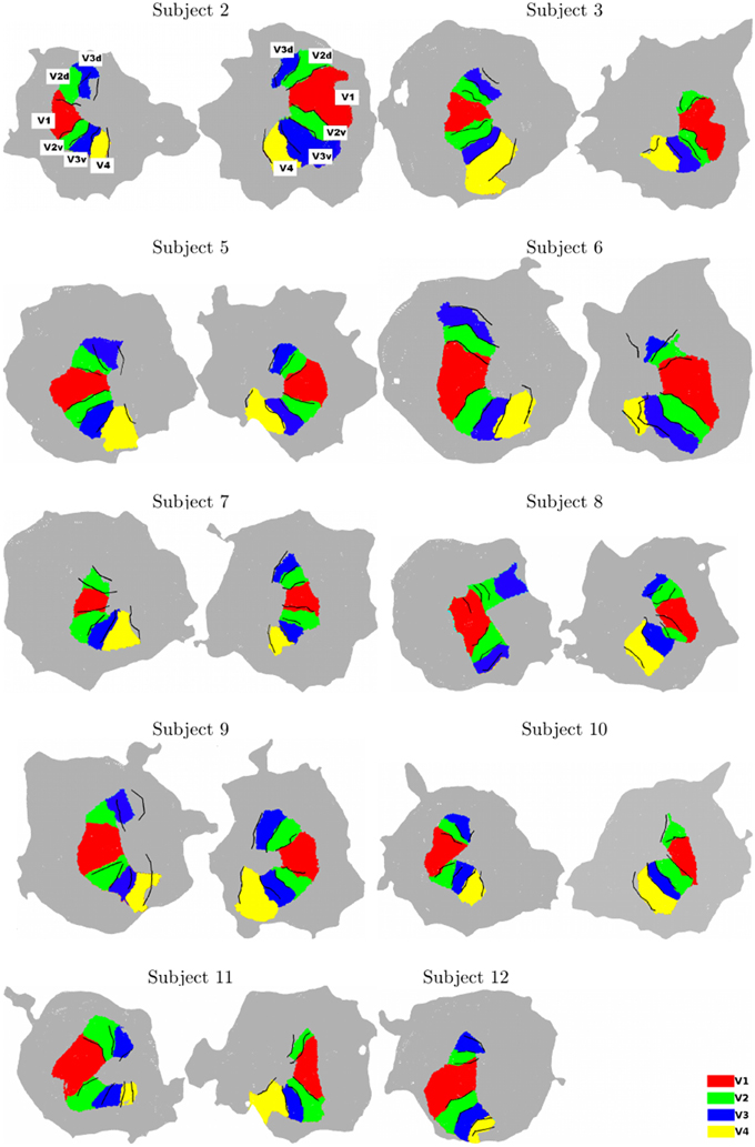
English: Surface models for some subjects (Left: left hemisphere, Right: right hemisphere, for each subject). The surface models and borders corresponding to each visual area were computed using software BALC (black lines). The surfaces of the visual areas (color patches) were computed using BrainVoyager and then projected on the BALC surface model. Some inconsistencies appear between the two methods in the dorsal part for two subjects (Subjects 8 and 9, left hemisphere) and BALC surface model failed for the right hemisphere of Subject 12. The visual area labels shown for Subject 2 are valid for all subjects. |
| Date | Published: 19 May 2015 |
| Source | Bordier C, Hupé J-M and Dojat M (2015) Quantitative evaluation of fMRI retinotopic maps, from V1 to V4, for cognitive experiments. Front. Hum. Neurosci. 9:277. doi: 10.3389/fnhum.2015.00277 http://journal.frontiersin.org/article/10.3389/fnhum.2015.00277/full |
| Author | Bordier C, Hupé J-M and Dojat M |
Licensing
[edit]
This file is licensed under the Creative Commons Attribution 4.0 International license.
- You are free:
- to share – to copy, distribute and transmit the work
- to remix – to adapt the work
- Under the following conditions:
- attribution – You must give appropriate credit, provide a link to the license, and indicate if changes were made. You may do so in any reasonable manner, but not in any way that suggests the licensor endorses you or your use.
File history
Click on a date/time to view the file as it appeared at that time.
| Date/Time | Thumbnail | Dimensions | User | Comment | |
|---|---|---|---|---|---|
| current | 20:37, 20 October 2015 | | 680 × 1,033 (372 KB) | Was a bee (talk | contribs) | == {{int:filedesc}} == {{Information |Description={{en|1=Surface models for some subjects (Left: left hemisphere, Right: right hemisphere, for each subject). The surface models and borders corresponding to each visual area were computed using software... |
You cannot overwrite this file.
File usage on Commons
There are no pages that use this file.