Category:Human visual cortex
Jump to navigation
Jump to search
see also
- Category:Human auditory cortex
- Category:Human somatosensory cortex
- Category:Human motor cortex
- Category:Language cortex
and
Subcategories
This category has the following 14 subcategories, out of 14 total.
- Videos of human visual cortex (19 F)
B
- Brodmann area 18 (16 F)
- Brodmann area 19 (14 F)
C
H
L
- Lunate sulcus (2 F)
M
- Mus musculus visual cortex (13 F)
O
- Occipital pole (6 F)
P
- Pericalcarine cortex (1 F)
R
- Retinotopy (13 F)
V
- Ventral and dorsal stream (16 F)
- Visual area MT (13 F)
- Visual area V4 (18 F)
Media in category "Human visual cortex"
The following 103 files are in this category, out of 103 total.
-
 1204 Optic Nerve vs Optic Tract fr.jpg 346 × 318; 21 KB
-
 Agrégat de cartes de champ visuel de Wandell et als 2007.png 472 × 839; 332 KB
-
 BA17 Primary visual cortex - animation.gif 300 × 300; 1.77 MB
-
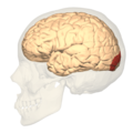
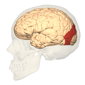
 BA17 Primary visual cortex - lateral view.png 1,200 × 1,200; 689 KB
-
 BA17 Primary visual cortex - medial view.png 1,200 × 1,200; 532 KB
-
 BA17 Primary visual cortex - posterior view.png 1,200 × 1,200; 436 KB
-
 BA17,18,19 - Visual cortex (V1, V2, V3) - animation.gif 300 × 300; 1.62 MB
-
 BA17,18,19 - Visual cortex (V1, V2, V3) - lateral view.png 1,200 × 1,200; 693 KB
-
 BA17,18,19 - Visual cortex (V1, V2, V3) - medial view.png 1,200 × 1,200; 533 KB
-
 BA17,18,19 - Visual cortex (V1, V2, V3) - posterior view.png 1,200 × 1,200; 441 KB
-
 BA18 - Secondary visual cortex (V2) - animation.gif 300 × 300; 1.73 MB
-
 BA18 - Secondary visual cortex (V2) - lateral view.png 1,200 × 1,200; 689 KB
-
 BA18 - Secondary visual cortex (V2) - medial view.png 1,200 × 1,200; 533 KB
-
 BA18 - Secondary visual cortex (V2) - posterior view.png 1,200 × 1,200; 438 KB
-
 BA19 - Visual association cortex (V3) - animation.gif 300 × 300; 1.72 MB
-
 BA19 - Visual association cortex (V3) - lateral view.png 1,200 × 1,200; 686 KB
-
 BA19 - Visual association cortex (V3) - medial view.png 1,200 × 1,200; 531 KB
-
 BA19 - Visual association cortex (V3) - posterior view.png 1,200 × 1,200; 435 KB
-
 Blausen 0103 Brain Sensory&Motor ar.png 1,600 × 960; 5.87 MB
-
 Blausen 0103 Brain Sensory&Motor.png 1,600 × 960; 993 KB
-
 Brain 2.jpg 721 × 540; 171 KB
-
 Brain image of blind echolocator.tif 1,200 × 923; 882 KB
-
 Brodmann areas 17 18 19.png 256 × 192; 40 KB
-
 Brodmann Cytoarchitectonics 17.png 599 × 952; 231 KB
-
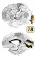 Brodmann Cytoarchitectonics 18.png 599 × 952; 225 KB
-
 Brodmann Cytoarchitectonics 19.png 599 × 952; 247 KB
-
 Cajal cortex drawings.png 672 × 777; 181 KB
-
 Candelabro 03 CUATRIS.jpg 377 × 445; 128 KB
-
 Carte retino cluster.png 886 × 531; 357 KB
-
 Caudal view of a brain after dissection.png 1,890 × 2,405; 2.94 MB
-
 Cerebral hemisphere of an ape. Wellcome L0001994.jpg 1,570 × 1,099; 566 KB
-
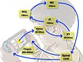
 Cerebro sistema.png 472 × 314; 57 KB
-
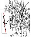
 Complex cell.JPG 214 × 196; 5 KB
-
 Constudeyepath ja.png 800 × 1,139; 112 KB
-
 Constudeyepath1.JPG 975 × 1,265; 441 KB
-
 Constudproc.png 763 × 865; 31 KB
-
 Cortex strie.svg 305 × 393; 39 KB
-
 Corticalorient.GIF 450 × 327; 5 KB
-
 Cytoarchitecture Visual cortex.jpg 709 × 809; 212 KB
-
 Dorsal and ventral attention systems.jpg 878 × 498; 106 KB
-
 Functional magnetic resonance imaging.jpg 250 × 208; 11 KB
-
 Fusiform face area face recognition.jpg 475 × 503; 95 KB
-
 Gray726 parieto-occipital sulcus.png 700 × 405; 86 KB
-
 Gray727 calcarine sulcus.svg 1,025 × 598; 22 KB
-
 Gray727 Cuneus.png 1,025 × 598; 144 KB
-
 Gray727 parieto-occipital fissure.svg 1,025 × 598; 22 KB
-
 Gray746.png 329 × 650; 21 KB
-
 Gray756.png 600 × 354; 32 KB
-
 Gray757.png 600 × 346; 27 KB
-

-
 Human visual cortex V1 cropped.png 900 × 349; 331 KB
-
 Human visual cortex V1.png 901 × 961; 820 KB
-
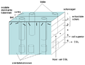
 Hypercolonne.png 927 × 596; 88 KB
-
 Hypercolonne.svg 294 × 230; 24 KB
-
 ImplantSawan.JPG 512 × 384; 15 KB
-
 Latency of response to a visual stimulus along the visual pathways.jpg 1,999 × 1,504; 393 KB
-
 Lateral and medial views of the marmoset cerebral cortex.jpg 2,667 × 793; 152 KB
-
 Lawrence 1960 9.3.png 1,784 × 2,792; 353 KB
-
 Lisa analysis.png 404 × 556; 136 KB
-
 Long-Evans rat V1 cortico-connection experiment - Fluorescent axon bundles in left V1.jpg 4,288 × 2,848; 4.56 MB
-
 Medial surface of cerebral cortex - ceneus.png 1,179 × 747; 514 KB
-
 Mod hierarchique Vasseur.svg 283 × 393; 26 KB
-
 Module.PNG 702 × 539; 34 KB
-
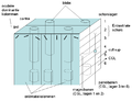 Module2.PNG 702 × 539; 35 KB
-
 Module3.PNG 702 × 539; 37 KB
-
 Neocórtex visual capas.png 305 × 600; 917 KB
-
 Neural Abstraction Pyramid.jpg 1,343 × 782; 253 KB
-
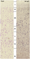
 Nissl-stained visual cortex of a human adult.png 419 × 777; 120 KB
-
 Optic nerve pair & two brain hemispheres.jpg 252 × 405; 21 KB
-
 Optic pathway.png 1,890 × 2,528; 1.29 MB
-
 Optical-transformations.png 1,415 × 2,085; 411 KB
-
 Parcellation of different cortical regions involved in visual processing.jpg 1,800 × 1,079; 205 KB
-
 Proj retine CGL cortex colonnes1.jpg 1,546 × 2,709; 326 KB
-
 Proj retine CGL cortex1.svg 549 × 857; 134 KB
-

-
 Schematic organization of visual cortex in the marmoset.jpg 2,667 × 2,289; 355 KB
-
 Scissure calca1.svg 354 × 461; 45 KB
-
 Sehbahn 3D v2.jpg 817 × 674; 94 KB
-
 Sehbahn 3D.jpg 817 × 674; 99 KB
-
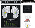 Sensitivity of human V6 to Translational egomotion.jpg 744 × 603; 231 KB
-
 Surface models and borders visual area.jpg 680 × 1,033; 372 KB
-
-
-
-
-
-
 V1 v2 v3.jpg 522 × 460; 32 KB
-
 Visual analisation.jpg 311 × 542; 48 KB
-
 Visual cortex - intermed mag.jpg 2,848 × 4,272; 8.74 MB
-
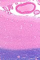 Visual cortex - low mag.jpg 2,848 × 4,272; 5.43 MB
-
 Visual cortex near fracture.png 819 × 784; 448 KB
-
 Visual cortex.jpg 589 × 432; 37 KB
-
 Visual field maps.jpg 886 × 886; 106 KB
-
 Visual Pathway to the Brain.jpg 960 × 720; 50 KB
-
 Visualcortex.gif 201 × 179; 10 KB
-
 VisualSingularity.jpg 599 × 393; 55 KB
-
 Visueel systeem.PNG 777 × 368; 17 KB
-
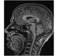
 Visueller Cortex(MRT).gif 393 × 320; 69 KB
-
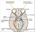
 Voies visuelles3.svg 744 × 964; 151 KB
-
 Voxel-man-brain.jpg 900 × 900; 497 KB
-
 Wandell maps.jpg 1,181 × 886; 114 KB
-
 Wat waar.PNG 616 × 443; 113 KB
-
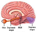 Зоровий шлях.jpg 250 × 218; 45 KB


_-_animation.gif)
_-_lateral_view.png)
_-_medial_view.png)
_-_posterior_view.png)
_-_animation.gif)
_-_lateral_view.png)
_-_medial_view.png)
_-_posterior_view.png)
_-_animation.gif)
_-_lateral_view.png)
_-_medial_view.png)
_-_posterior_view.png)


.gif)


