File:Structural basis for DNA binding by the PolD–PCNA complex.pdf
Jump to navigation
Jump to search

Size of this JPG preview of this PDF file: 800 × 474 pixels. Other resolutions: 320 × 190 pixels | 640 × 379 pixels.
Original file (1,012 × 600 pixels, file size: 1.4 MB, MIME type: application/pdf)
Captions
Captions
Structural basis for DNA binding by the PolD–PCNA complex
Summary
[edit]| Description |
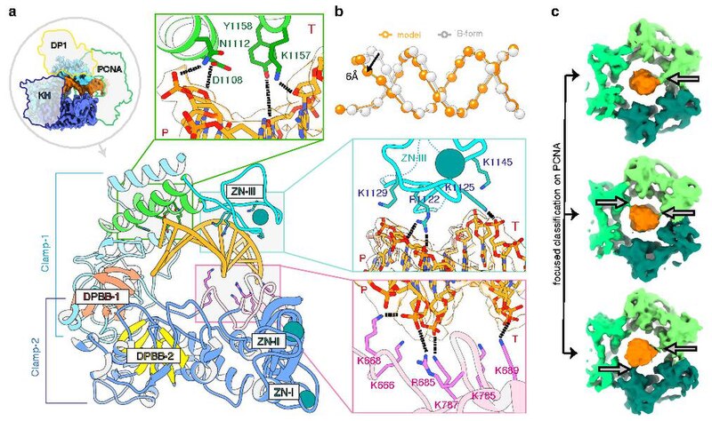
English: a View of the DP2 clamp-1 (light blue) and clamp-2 (dark blue) domains surrounding one turn of the DNA duplex (orange). The two catalytic double-psi β-barrels DPBB-1 and DPBB-2 are represented in red and yellow, respectively. Template and primer strands are indicated by T and P, respectively. b Interaction with the PolD clamp causes a local distortion of the DNA (orange) compared with an ideal B-form DNA (white). The phosphates of the DNA duplexes are shown as spheres. c PCNA makes labile contacts with DNA. Focused classification on PCNA resulted in three 3D classes showing extra-densities between DNA and PCNA residues K84 and K86, which are underlined by arrows.[1] |
| Date | |
| Source | https://doi.org/10.1038/s41467-020-15392-9 |
| Author | Clément Madru, Ghislaine Henneke, Pierre Raia, Inès Hugonneau-Beaufet, Gérard Pehau-Arnaudet, Patrick England, Erik Lindahl, Marc Delarue, Marta Carroni & Ludovic Sauguet |
Licensing
[edit]This file is licensed under the Creative Commons Attribution 4.0 International license.
- You are free:
- to share – to copy, distribute and transmit the work
- to remix – to adapt the work
- Under the following conditions:
- attribution – You must give appropriate credit, provide a link to the license, and indicate if changes were made. You may do so in any reasonable manner, but not in any way that suggests the licensor endorses you or your use.
File history
Click on a date/time to view the file as it appeared at that time.
| Date/Time | Thumbnail | Dimensions | User | Comment | |
|---|---|---|---|---|---|
| current | 23:32, 4 April 2020 |  | 1,012 × 600 (1.4 MB) | Rob Hurt (talk | contribs) | Uploaded a work by Clément Madru, Ghislaine Henneke, Pierre Raia, Inès Hugonneau-Beaufet, Gérard Pehau-Arnaudet, Patrick England, Erik Lindahl, Marc Delarue, Marta Carroni & Ludovic Sauguet from https://doi.org/10.1038/s41467-020-15392-9 with UploadWizard |
You cannot overwrite this file.
File usage on Commons
There are no pages that use this file.
File usage on other wikis
The following other wikis use this file: