File:SEM for a Glioblstoma Cell Exposed to CeO2.tif
Jump to navigation
Jump to search

Size of this JPG preview of this TIF file: 800 × 545 pixels. Other resolutions: 320 × 218 pixels | 640 × 436 pixels | 1,024 × 698 pixels | 1,280 × 872 pixels | 2,560 × 1,744 pixels | 4,037 × 2,750 pixels.
Original file (4,037 × 2,750 pixels, file size: 10.59 MB, MIME type: image/tiff)
Captions
Captions
Add a one-line explanation of what this file represents
Summary
[edit]| Description |
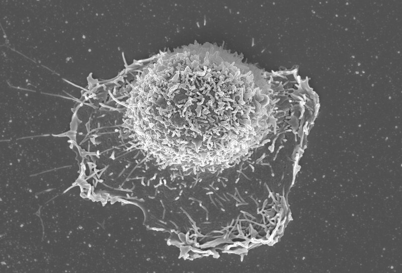
English: SEM for a single glioblastoma cell after treatment with cerium oxide nanoparticles. The image illustrates a swelling cell, with an induction of the microvillus formation. It was captured using ZEISS SEM at KAUST core labs, with a magnification of 9000X. |
| Date | |
| Source | Own work |
| Author | Bader Aloufi |
Licensing
[edit]I, the copyright holder of this work, hereby publish it under the following license:
This file is licensed under the Creative Commons Attribution 4.0 International license.
- You are free:
- to share – to copy, distribute and transmit the work
- to remix – to adapt the work
- Under the following conditions:
- attribution – You must give appropriate credit, provide a link to the license, and indicate if changes were made. You may do so in any reasonable manner, but not in any way that suggests the licensor endorses you or your use.
| This image was uploaded as part of Wiki Science Competition 2017. |
File history
Click on a date/time to view the file as it appeared at that time.
| Date/Time | Thumbnail | Dimensions | User | Comment | |
|---|---|---|---|---|---|
| current | 15:09, 15 December 2017 |  | 4,037 × 2,750 (10.59 MB) | Bader Aloufi (talk | contribs) | User created page with UploadWizard |
You cannot overwrite this file.
File usage on Commons
The following 2 pages use this file: