File:Rapid-and-Efficient-Clearance-of-Blood-borne-Virus-by-Liver-Sinusoidal-Endothelium-ppat.1002281.s007.ogv
Jump to navigation
Jump to search
Size of this JPG preview of this OGG file: 600 × 600 pixels. Other resolutions: 240 × 240 pixels | 480 × 480 pixels | 640 × 640 pixels.
Original file (Ogg Theora video file, length 1.6 s, 640 × 640 pixels, 3.6 Mbps, file size: 703 KB)
Captions
Captions
Add a one-line explanation of what this file represents
Summary
[edit]| Description |
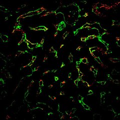
English: Virus (red) + LSEC (pseudo-green). (Quick time; 3 MB MB) The great majority of red virus is either in, on, or very near the green LSEC image suggesting close association of the two. Details of image acquisition and 3D video creation are given in Materials and Methods. |
||
| Date | |||
| Source | Video S4 from Ganesan L, Mohanty S, Kim J, Clark K, Robinson J, Anderson C (2011). "Rapid and Efficient Clearance of Blood-borne Virus by Liver Sinusoidal Endothelium". PLOS Pathogens. DOI:10.1371/journal.ppat.1002281. PMID 21980295. PMC: 3182912. | ||
| Author | Ganesan L, Mohanty S, Kim J, Clark K, Robinson J, Anderson C | ||
| Permission (Reusing this file) |
|
||
| Provenance |
|
File history
Click on a date/time to view the file as it appeared at that time.
| Date/Time | Thumbnail | Dimensions | User | Comment | |
|---|---|---|---|---|---|
| current | 04:06, 21 November 2012 | 1.6 s, 640 × 640 (703 KB) | Open Access Media Importer Bot (talk | contribs) | Automatically uploaded media file from Open Access source. Please report problems or suggestions here. |
You cannot overwrite this file.
File usage on Commons
There are no pages that use this file.
Transcode status
Update transcode statusMetadata
Categories:
- Immunologic techniques
- Immunohistochemical analysis
- Videos of leukocytes
- Immune response
- Viral clearance
- Viral vectors
- Videos of virus infection in endothelial cells
- Infectious diseases in Mus musculus
- Immunodeficiency viruses
- Viral immune evasion
- Adenoviridae infections
- Blood-borne pathogens
- Vascular endothelium
- Kupffer cells
- Inbred BALB C mice