File:NUP-1-Is-a-Large-Coiled-Coil-Nucleoskeletal-Protein-in-Trypanosomes-with-Lamin-Like-Functions-pbio.1001287.s007.ogv
Jump to navigation
Jump to search
Size of this JPG preview of this OGG file: 600 × 600 pixels. Other resolutions: 240 × 240 pixels | 480 × 480 pixels | 768 × 768 pixels | 1,024 × 1,024 pixels | 2,048 × 2,048 pixels.
Original file (Ogg Theora video file, length 9.0 s, 2,048 × 2,048 pixels, 149 kbps, file size: 163 KB)
Captions
Captions
Add a one-line explanation of what this file represents
Summary
[edit]| Description |
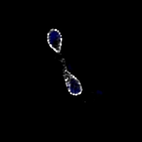
English: NUP-1 forms a network-like structure around the periphery of the nucleus. PCF cells expressing NUP-1-GFP (white) were imaged by confocal microscopy. Shown is an image series along the z-axis of a cell during nuclear division. DAPI is used to visualize DNA (blue). |
||
| Date | |||
| Source | Movie S2 from DuBois K, Alsford S, Holden J, Buisson J, Swiderski M, Bart J, Ratushny A, Wan Y, Bastin P, Barry J, Navarro M, Horn D, Aitchison J, Rout M, Field M (2012). "NUP-1 Is a Large Coiled-Coil Nucleoskeletal Protein in Trypanosomes with Lamin-Like Functions". PLOS Biology. DOI:10.1371/journal.pbio.1001287. PMID 22479148. PMC: 3313915. | ||
| Author | DuBois K, Alsford S, Holden J, Buisson J, Swiderski M, Bart J, Ratushny A, Wan Y, Bastin P, Barry J, Navarro M, Horn D, Aitchison J, Rout M, Field M | ||
| Permission (Reusing this file) |
|
||
| Provenance |
|
File history
Click on a date/time to view the file as it appeared at that time.
| Date/Time | Thumbnail | Dimensions | User | Comment | |
|---|---|---|---|---|---|
| current | 02:25, 31 October 2012 | 9.0 s, 2,048 × 2,048 (163 KB) | Open Access Media Importer Bot (talk | contribs) | Automatically uploaded media file from Open Access source. Please report problems or suggestions here. |
You cannot overwrite this file.
File usage on Commons
There are no pages that use this file.
Transcode status
Update transcode statusMetadata
Categories:
- Antigenic variation
- Cell nucleus
- Videos of chromosomes
- Gene expression regulation
- Gene knockdown techniques
- Protozoan genes
- Genetic loci
- Heterochromatin
- Lamins
- Transmission electron microscope videos
- Nuclear envelope
- Nuclear pore complex proteins
- Videos of nuclear proteins
- Protein conformation
- Protein transport
- Protozoan proteins
- Telomeres
- Transcription (genetics)
- Trypanosoma brucei brucei
- Trypanosoma variant surface glycoproteins