File:Lymphatic vessels develop in the postnatal ovary from around 10 dpn.png

Original file (3,439 × 2,585 pixels, file size: 4.46 MB, MIME type: image/png)
Captions
Captions
Summary
[edit]
| Description |
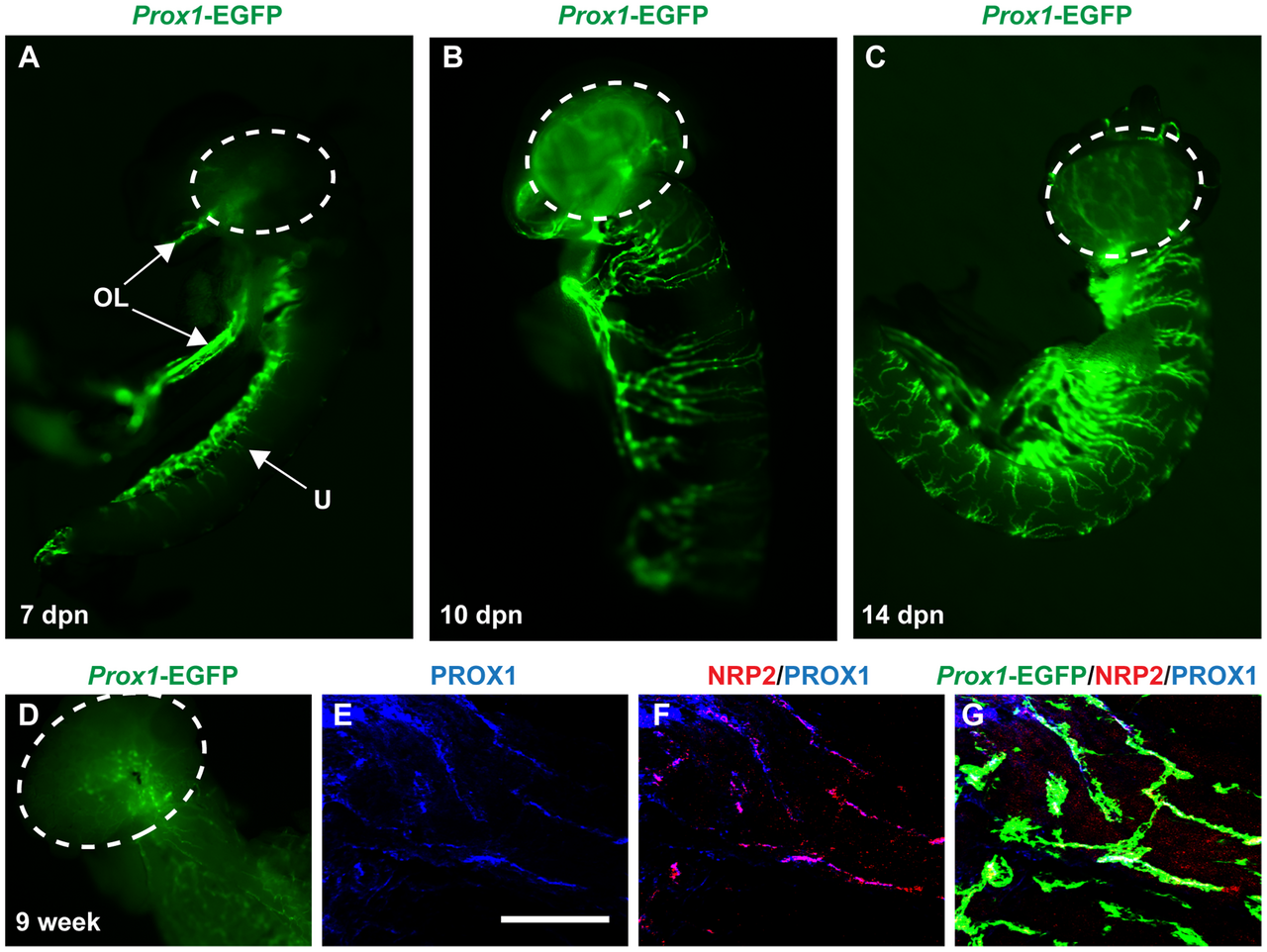
Figure 4. Lymphatic vessels develop in the postnatal ovary from around 10 dpn. A) At 7 dpn, EGFP-positive vessels are observed in along one side of the uterine horn and in the ovarian ligaments, but the ovary (encircled) is still devoid of lymphatics. B) At 10 dpn, the ovary possesses a lymphatic network. Lymphatic vessels sprouting laterally at distinct regional distances from a pre-existing vasculature along the length of the uterus have almost encircled the entire uterine horn. C) At 14 dpn, the ovary possesses a distinct lymphatic network and the uterine horn has developed a strikingly segmented lymphatic network encircling the entire tissue D) The adult ovary maintain a high Prox1-RGFP expression and the uterine horn has developed a extensive mesh of lymphatic vessels. E) Prominent Prox1-EGFP positive vessels of the 9 week uterine horn also express endogenous PROX1, and F) the lymphatic marker NRP2, G) both overlapping with Prox1-EGFP expression. OL = ovarian ligament; U = uterine horn; scale bar = 200 µm. https://doi.org/10.1371/journal.pone.0052620.g004 |
| Date | |
| Source | https://journals.plos.org/plosone/article?id=10.1371/journal.pone.0052620 Svingen T, François M, Wilhelm D, Koopman P (2012) Three-Dimensional Imaging of Prox1-EGFP Transgenic Mouse Gonads Reveals Divergent Modes of Lymphangiogenesis in the Testis and Ovary. PLoS ONE 7(12): e52620. https://doi.org/10.1371/journal.pone.0052620 |
| Author | Lim J, Maher GJ, Turner GDH, Dudka-Ruszkowska W, Taylor S, Meyts ER-D, et al. |
Licensing
[edit]
© 2012 Svingen et al. This is an open-access article distributed under the terms of the Creative Commons Attribution License, which permits unrestricted use, distribution, and reproduction in any medium, provided the original author and source are credited.

|
This file, which was originally posted to an external website, has not yet been reviewed by an administrator or reviewer to confirm that the above license is valid. See Category:License review needed for further instructions.
|
- You are free:
- to share – to copy, distribute and transmit the work
- to remix – to adapt the work
- Under the following conditions:
- attribution – You must give appropriate credit, provide a link to the license, and indicate if changes were made. You may do so in any reasonable manner, but not in any way that suggests the licensor endorses you or your use.
File history
Click on a date/time to view the file as it appeared at that time.
| Date/Time | Thumbnail | Dimensions | User | Comment | |
|---|---|---|---|---|---|
| current | 20:23, 2 August 2024 | | 3,439 × 2,585 (4.46 MB) | Rasbak (talk | contribs) | {{Information |description=Figure 4. Lymphatic vessels develop in the postnatal ovary from around 10 dpn. A) At 7 dpn, EGFP-positive vessels are observed in along one side of the uterine horn and in the ovarian ligaments, but the ovary (encircled) is still devoid of lymphatics. B) At 10 dpn, the ovary possesses a lymphatic network. Lymphatic vessels sprouting laterally at distinct regional distances from a pre-existing vasculature along the length of the uterus have almost encircled the enti... |
You cannot overwrite this file.
File usage on Commons
There are no pages that use this file.