Category:Rete testis
Jump to navigation
Jump to search
Subcategories
This category has the following 3 subcategories, out of 3 total.
A
H
Media in category "Rete testis"
The following 27 files are in this category, out of 27 total.
-
 3D reconstruction of immunopositive tubules.png 2,098 × 1,376; 2.64 MB
-

-
 An American text-book of physiology (1897) (14780995834).jpg 838 × 1,504; 200 KB
-
 Anatomical description of testicular and epididymal structures.jpg 1,362 × 960; 641 KB
-
 Cystic dysplasia of rete testis.jpg 677 × 469; 426 KB
-
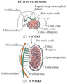 Desarrollo Testicular.png 453 × 564; 155 KB
-
 EB1911 Reproductive System, in Anatomy - testis and epididymis.jpg 408 × 414; 87 KB
-
 Embryonic development of the mouse rete testis.png 1,497 × 1,257; 1.24 MB
-
 Figure 28 01 03.jpg 743 × 686; 367 KB
-
 Gray1149.png 609 × 891; 547 KB
-
 Hodenschema.svg 744 × 1,052; 57 KB
-
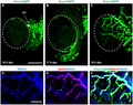
 Lymphatic vessels are limited to the tunica albuginea in adult testis.png 3,439 × 2,951; 11.7 MB
-
 Lymphatic vessels develop in the postnatal ovary from around 10 dpn.png 3,439 × 2,585; 4.46 MB
-
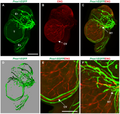 Lymphatic vessels sprout across, but not beyond, the testis cap at 17.5 dpc.png 3,439 × 3,257; 8.81 MB
-
 Morphology of the human testis. A. Cross section of the human testes.png 850 × 467; 604 KB
-

-
 Rete testis.jpg 1,663 × 1,833; 1.06 MB
-

-

-

-
 Structure and organisation of the human testis and seminiferous tubules.png 2,098 × 845; 860 KB
-
 Testicle-Revised.jpg 1,663 × 1,833; 1.42 MB
-
-

-
 The adult ovary possesses an extensive lymphatic network.png 3,439 × 2,968; 14.27 MB
-

-
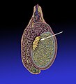
 Ultrasound-guided injection of the rete testis of a monkey.png 1,048 × 1,193; 1.1 MB

_(14784000323).jpg)
_(14780995834).jpg)


,_juvenile_(18_M),_and_adult_(60_M)_rhesus_monkey_testes.png)


.jpg)