File:Ion-Abrasion-Scanning-Electron-Microscopy-Reveals-Surface-Connected-Tubular-Conduits-in-HIV-ppat.1000591.s009.ogv
Jump to navigation
Jump to search
Size of this JPG preview of this OGG file: 695 × 600 pixels. Other resolutions: 278 × 240 pixels | 556 × 480 pixels | 890 × 768 pixels | 1,024 × 884 pixels.
Original file (Ogg Theora video file, length 3.4 s, 1,024 × 884 pixels, 11.98 Mbps, file size: 4.9 MB)
Captions
Captions
Add a one-line explanation of what this file represents
Summary
[edit]| Description |
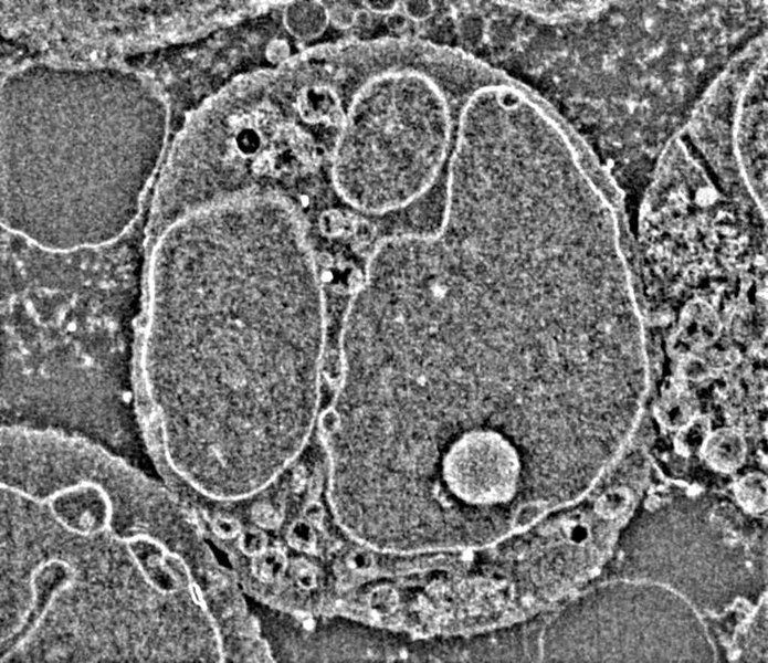
English: Example of image stacks obtained by IA-SEM illustrating the interiors of Jurkat T cells infected with the 29/31 Gag matrix mutant. The cross-sectional views include the width of the entire cell, and show numerous spherical vacuoles, likely to represent endosomal compartments. There is no evidence of long virion channels in these cells. |
||
| Date | |||
| Source | Video S9 from Bennett A, Narayan K, Shi D, Hartnell L, Gousset K, He H, Lowekamp B, Yoo T, Bliss D, Freed E, Subramaniam S (2009). "Ion-Abrasion Scanning Electron Microscopy Reveals Surface-Connected Tubular Conduits in HIV-Infected Macrophages". PLOS Pathogens. DOI:10.1371/journal.ppat.1000591. PMID 19779568. PMC: 2743285. | ||
| Author | Bennett A, Narayan K, Shi D, Hartnell L, Gousset K, He H, Lowekamp B, Yoo T, Bliss D, Freed E, Subramaniam S | ||
| Permission (Reusing this file) |
|
||
| Provenance |
|
File history
Click on a date/time to view the file as it appeared at that time.
| Date/Time | Thumbnail | Dimensions | User | Comment | |
|---|---|---|---|---|---|
| current | 13:05, 17 November 2012 | 3.4 s, 1,024 × 884 (4.9 MB) | Open Access Media Importer Bot (talk | contribs) | Automatically uploaded media file from Open Access source. Please report problems or suggestions here. |
You cannot overwrite this file.
File usage on Commons
There are no pages that use this file.