File:High-Resolution-Intravital-Microscopy-pone.0050915.s005.ogv
Jump to navigation
Jump to search
Size of this JPG preview of this OGG file: 431 × 600 pixels. Other resolutions: 172 × 239 pixels | 468 × 651 pixels.
Original file (Ogg Theora video file, length 3.8 s, 468 × 651 pixels, 573 kbps, file size: 266 KB)
Captions
Captions
Add a one-line explanation of what this file represents
Summary
[edit]| Description |
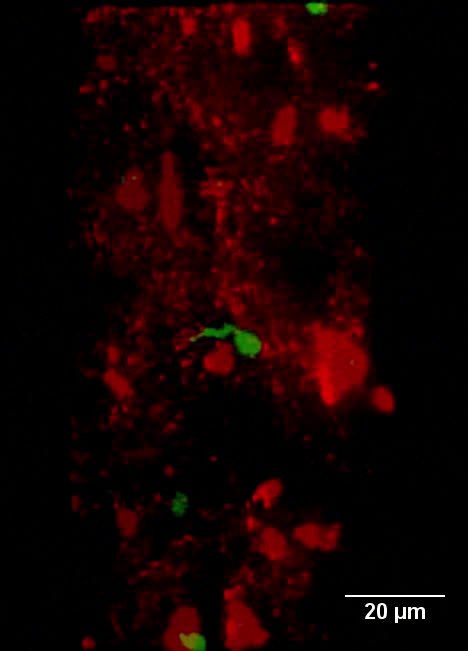
English: Rotation view of a 3D fluorescence image (465×651×11 voxel, 186×260×10 µm3) in a germinal center of the popliteal lymph node of a NP-CGG immunized mouse, 9 days after immunization. B1–8+/+ Jκ−/− EGFP+/+ cells previously transferred to the C57BL6 mouse are represented in green and follicular dendritic cells (FDCs) in situ stained with anti-CD21/35-Fab fragment-Alexa568 are represented in red. λexc = 800 nm. |
||
| Date | |||
| Source | Movie S1 from Andresen V, Pollok K, Rinnenthal J, Oehme L, Günther R, Spiecker H, Radbruch H, Gerhard J, Sporbert A, Cseresnyes Z, Hauser A, Niesner R (2012). "High-Resolution Intravital Microscopy". PLOS ONE. DOI:10.1371/journal.pone.0050915. PMID 23251402. PMC: 3522675. | ||
| Author | Andresen V, Pollok K, Rinnenthal J, Oehme L, Günther R, Spiecker H, Radbruch H, Gerhard J, Sporbert A, Cseresnyes Z, Hauser A, Niesner R | ||
| Permission (Reusing this file) |
This file is licensed under the Creative Commons Attribution 3.0 Unported license.
|
||
| Provenance |
|
File history
Click on a date/time to view the file as it appeared at that time.
| Date/Time | Thumbnail | Dimensions | User | Comment | |
|---|---|---|---|---|---|
| current | 15:07, 15 October 2016 | 3.8 s, 468 × 651 (266 KB) | Open Access Media Importer Bot (talk | contribs) | Automatically uploaded media file from Open Access source. Please report problems or suggestions here. |
You cannot overwrite this file.
File usage on Commons
There are no pages that use this file.
Transcode status
Update transcode statusMetadata
Categories:
- Videos of leukocytes
- Videos of B cells
- Immune response
- Immunologic techniques
- Animal models of disease
- Videos of signal processing
- Videos of image processing
- Videos of electromagnetic radiation
- Interdisciplinary physics
- Videos of immune system
- Confocal microscopic movies from Open Access Journals
- Media from PLOS ONE