File:Experimental-Cerebral-Malaria-Pathogenesis—Hemodynamics-at-the-Blood-Brain-Barrier-ppat.1004528.s031.ogv
Jump to navigation
Jump to search
Size of this JPG preview of this OGG file: 571 × 600 pixels. Other resolutions: 229 × 240 pixels | 457 × 480 pixels | 679 × 713 pixels.
Original file (Ogg Theora video file, length 4.0 s, 679 × 713 pixels, 1.82 Mbps, file size: 887 KB)
Captions
Captions
Add a one-line explanation of what this file represents
Summary
[edit]| Description |
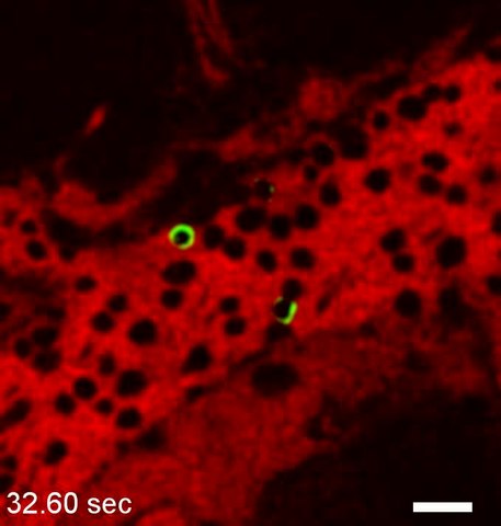
English: CD4+ T cell behavior during ECM. Intravital microscopy of a PbA infected CBA/CaJ mouse with ECM showing CD4+ T cells (green surface label) arrested on the endothelium of a cortical postcapillary venules. CD4+ T cells were labeled by intravenous inoculation of PE-conjugated anti-CD4+. The vascular lumen was visualized with Evans blue (red). The numerous large leukocytes (dark circles) represent most likely monocytes and macrophages. Scale bar = 20 µm. |
||
| Date | |||
| Source | Video S8 from Nacer A, Movila A, Sohet F, Girgis N, Gundra U, Loke P, Daneman R, Frevert U (2014). "Experimental Cerebral Malaria Pathogenesis—Hemodynamics at the Blood Brain Barrier". PLOS Pathogens. DOI:10.1371/journal.ppat.1004528. PMID 25474413. PMC: 4256476. | ||
| Author | Nacer A, Movila A, Sohet F, Girgis N, Gundra U, Loke P, Daneman R, Frevert U | ||
| Permission (Reusing this file) |
This file is licensed under the Creative Commons Attribution 4.0 International license.
|
||
| Provenance |
|
File history
Click on a date/time to view the file as it appeared at that time.
| Date/Time | Thumbnail | Dimensions | User | Comment | |
|---|---|---|---|---|---|
| current | 22:51, 13 December 2014 | 4.0 s, 679 × 713 (887 KB) | Open Access Media Importer Bot (talk | contribs) | Automatically uploaded media file from Open Access source. Please report problems or suggestions here. |
You cannot overwrite this file.
File usage on Commons
There are no pages that use this file.