File:Endothelial-Cell-Self-fusion-during-Vascular-Pruning-pbio.1002126.s009.ogv
Jump to navigation
Jump to search
Size of this JPG preview of this OGG file: 800 × 559 pixels. Other resolutions: 320 × 224 pixels | 640 × 448 pixels | 1,024 × 716 pixels | 1,280 × 895 pixels | 1,716 × 1,200 pixels.
Original file (Ogg Theora video file, length 4.5 s, 1,716 × 1,200 pixels, 7.51 Mbps, file size: 4.03 MB)
Captions
Captions
Add a one-line explanation of what this file represents
Summary
[edit]| Description |
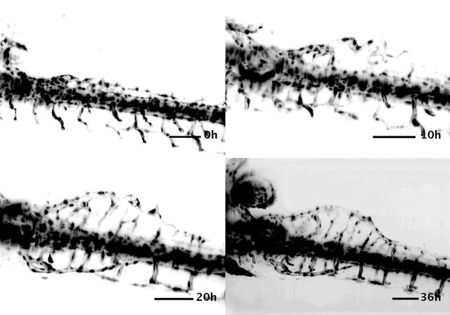
English: Key phases of SIV development—3-D projection of mSPIM images. Related to Fig 1A–1E”. Images are projections of 3-D SPIM images extracted from a time-lapse movie, showing four key phases of SIV development at ~36, 46, 56, and 72 hpf. The stages shown correspond to models in Fig 1A–1D. The movie shows a 360° turn around the anterior-posterior axis, showing SIV plexuses on both sides of the embryo. |
||
| Date | |||
| Source | S2 Movie from Lenard A, Daetwyler S, Betz C, Ellertsdottir E, Belting H, Huisken J, Affolter M (2015). "Endothelial Cell Self-fusion during Vascular Pruning". PLOS Biology. DOI:10.1371/journal.pbio.1002126. PMID 25884426. PMC: 4401649. | ||
| Author | Lenard A, Daetwyler S, Betz C, Ellertsdottir E, Belting H, Huisken J, Affolter M | ||
| Permission (Reusing this file) |
This file is licensed under the Creative Commons Attribution 4.0 International license.
|
||
| Provenance |
|
File history
Click on a date/time to view the file as it appeared at that time.
| Date/Time | Thumbnail | Dimensions | User | Comment | |
|---|---|---|---|---|---|
| current | 23:49, 23 April 2015 | 4.5 s, 1,716 × 1,200 (4.03 MB) | Open Access Media Importer Bot (talk | contribs) | Automatically uploaded media file from Open Access source. Please report problems or suggestions here. |
You cannot overwrite this file.
File usage on Commons
The following 2 pages use this file: