File:Dual-Role-of-Topoisomerase-II-in-Centromere-Resolution-and-Aurora-B-Activity-pbio.0060207.sv006.ogv
Jump to navigation
Jump to search
Size of this JPG preview of this OGG file: 800 × 354 pixels. Other resolutions: 320 × 141 pixels | 1,020 × 451 pixels.
Original file (Ogg Theora video file, length 4.0 s, 1,020 × 451 pixels, 358 kbps, file size: 175 KB)
Captions
Captions
Add a one-line explanation of what this file represents
Summary
[edit]| Description |
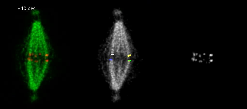
English: Time-Lapse Microscopy of S2 Cells Stably Expressing GFP-α-Tubulin and CID-mCherry That Were Depleted for TOPO II, 72 h after the Addition of the dsRNA Z-stacks were acquired every 40 s. Each Z-stack is 10 μm and is composed of ten optical sections. Anaphase onset corresponds to time 0 s. Merged color images for Cherry-CID (red) and GFP-α-tubulin (green) are shown on the left. In the middle, the separated channel for GFP-α-tubulin (white) is shown with representation of the CID pairs composing each centromere (on the right, separated channel). |
||
| Date | |||
| Source | Video S6 from Coelho P, Queiroz-Machado J, Carmo A, Moutinho-Pereira S, Maiato H, Sunkel C (2008). "Dual Role of Topoisomerase II in Centromere Resolution and Aurora B Activity". PLOS Biology. DOI:10.1371/journal.pbio.0060207. PMID 18752348. PMC: 2525683. | ||
| Author | Coelho P, Queiroz-Machado J, Carmo A, Moutinho-Pereira S, Maiato H, Sunkel C | ||
| Permission (Reusing this file) |
|
||
| Provenance |
|
File history
Click on a date/time to view the file as it appeared at that time.
| Date/Time | Thumbnail | Dimensions | User | Comment | |
|---|---|---|---|---|---|
| current | 23:25, 30 October 2012 | 4.0 s, 1,020 × 451 (175 KB) | Open Access Media Importer Bot (talk | contribs) | Automatically uploaded media file from Open Access source. Please report problems or suggestions here. |
You cannot overwrite this file.
File usage on Commons
There are no pages that use this file.
Transcode status
Update transcode statusMetadata
Categories:
- Videos of 2008
- Cell cycle proteins
- Videos of cultured cells
- Centromere
- Non-histone chromosomal proteins
- Chromosome segregation
- Type II DNA topoisomerases
- Videos of Drosophila melanogaster proteins
- Enzyme activation
- Schneider 2 cells
- Kinetochores
- Videos of microtubules of the spindle apparatus
- Protein-serine-threonine kinases
- RNA interference
- Sister chromatid exchange
- Topoisomerase II inhibitors
- MCherry
- Aurora kinase B