File:Coupling-Mechanical-Deformations-and-Planar-Cell-Polarity-to-Create-Regular-Patterns-in-the-pcbi.1002618.s010.ogv
From Wikimedia Commons, the free media repository
Jump to navigation
Jump to search
Size of this JPG preview of this OGG file: 383 × 599 pixels. Other resolutions: 153 × 239 pixels | 513 × 802 pixels.
Original file (Ogg Theora video file, length 25 s, 513 × 802 pixels, 3.83 Mbps, file size: 11.23 MB)
File information
Structured data
Captions
Captions
Add a one-line explanation of what this file represents
Summary
[edit]DescriptionCoupling-Mechanical-Deformations-and-Planar-Cell-Polarity-to-Create-Regular-Patterns-in-the-pcbi.1002618.s010.ogv |
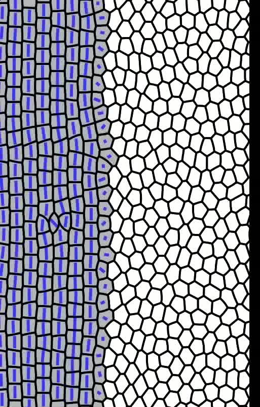
English: Simulation of the progressive growth of a cone photoreceptor mosaic similar to that observed near the retinal margin in adult, wildtype fish; the cone fate is induced and the PCP pathway is activated in successive columns of cells at regular intervals, as described in the text. |
||
Date | |||
Source | Video S1 from Salbreux G, Barthel L, Raymond P, Lubensky D. "Coupling Mechanical Deformations and Planar Cell Polarity to Create Regular Patterns in the Zebrafish Retina". PLOS Computational Biology. DOI:10.1371/journal.pcbi.1002618. PMID 22936893. PMC: 3426565. | ||
Author | Salbreux G, Barthel L, Raymond P, Lubensky D | ||
Permission (Reusing this file) |
|
||
Provenance InfoField |
|
File history
Click on a date/time to view the file as it appeared at that time.
Date/Time | Thumbnail | Dimensions | User | Comment | |
---|---|---|---|---|---|
current | 09:40, 19 October 2012 | 25 s, 513 × 802 (11.23 MB) | Open Access Media Importer Bot (talk | contribs) | Uploaded with the Open Access Media Importer. (test edit) botrequest |
You cannot overwrite this file.
File usage on Commons
There are no pages that use this file.
Transcode status
Update transcode statusMetadata
This file contains additional information such as Exif metadata which may have been added by the digital camera, scanner, or software program used to create or digitize it. If the file has been modified from its original state, some details such as the timestamp may not fully reflect those of the original file. The timestamp is only as accurate as the clock in the camera, and it may be completely wrong.
Author | Salbreux G, Barthel L, Raymond P, Lubensky D |
---|---|
Usage terms | http://creativecommons.org/licenses/by/3.0/ |
Image title | Simulation of the progressive growth of a cone photoreceptor mosaic similar to that observed near the retinal margin in adult, wildtype fish; the cone fate is induced and the PCP pathway is activated in successive columns of cells at regular intervals, as described in the text. |
Software used | Xiph.Org libtheora 1.1 20090822 (Thusnelda) |
Date and time of digitizing | 2012-08-23 |
Structured data
Items portrayed in this file
depicts
23 August 2012
application/ogg
331cf05298542561834479a7fce6e5076164a9a5
11,777,503 byte
24.6 second
802 pixel
513 pixel
Hidden categories:
- CC-BY-2.5
- Uploaded with Open Access Media Importer
- Media needing category review as of 21 October 2012
- Media needing category review with 2 suggested categories
- Uploaded with Open Access Media Importer and needing category review
- Media from PLOS Computational Biology
- Videos from open-access scholarly articles
- Ogv videos
- Video display resolution 513 x 802