File:Cisti aneurismatica.jpg
Jump to navigation
Jump to search
No higher resolution available.
Cisti_aneurismatica.jpg (650 × 450 pixels, file size: 52 KB, MIME type: image/jpeg)
Captions
Captions
Add a one-line explanation of what this file represents
Summary
[edit]
| Description |
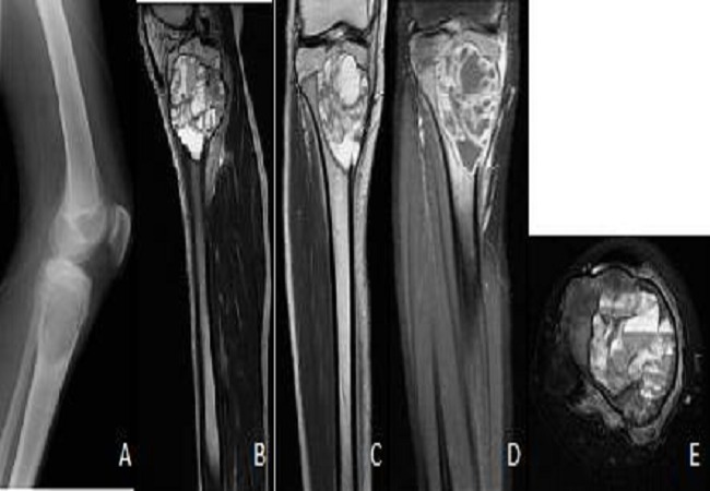
Italiano: Cisti aneurismatica localizzata a livello della porzione prossimale della tibia vista mediante radiografia (A) e mediante RM con sequenze pesate in T1(B), T2(C) T1 dopo somministrazione di mezzo di contrasto (D)e T2 FATSAT
English: Radiography (A) showing an osteolytic lesion oval metaphyseal tibial very limited.
Cuts MRI sagittal T1-weighted (B), coronal T2 (C) and T1 post contrast (D) and axial T2 FATSAT (E) showing the partitions after intralesional contrast enhanced liquid level typical of an aneurysmal cyst. |
| Date | |
| Source | Meryem Boubbou et al. 2013. Aneurysmal bone cyst primary - about eight pediatric cases: radiological aspects and review of the literature. The Pan African Medical Journal. |
| Author | Meryem Boubbou, Karima Atarraf, Lamiae Chater, Abderrahmane Afifi, Siham Tizniti |
| Permission (Reusing this file) |
CC-BY-2.0. |
Licensing
[edit]
This file is licensed under the Creative Commons Attribution 2.0 Generic license.
- You are free:
- to share – to copy, distribute and transmit the work
- to remix – to adapt the work
- Under the following conditions:
- attribution – You must give appropriate credit, provide a link to the license, and indicate if changes were made. You may do so in any reasonable manner, but not in any way that suggests the licensor endorses you or your use.
This file, which was originally posted to
The Pan African Medical Journal, was reviewed on 7 January 2019 by reviewer Ruthven, who confirmed that it was available there under the stated license on that date.
|
Original upload log
[edit]
The original description page was here. All following user names refer to it.wikipedia.
| Date/Time | Dimensions | User | Comment |
|---|---|---|---|
| 2019-01-07 14:03 | 650×450× (53093 bytes) | Galati Antonello | Cisti aneurismatica localizzata a livello della porzione prossimale della tibia vista mediante radiografia (A) e mediante RM con sequenze pesate in T1(B), T2(C) T1 dopo somministrazione di mezzo di contrasto (D)e T2 FATSAT Immagine tratta da Meryem Boubbou et al. 2013 Aneurysmal bone cyst primary - about eight pediatric cases: radiological aspects and review of the literature. The Pan African Medical Journal. CC-BY licence http://www.panafrican-med-journal.com/content/article/15/111/full/ |
File history
Click on a date/time to view the file as it appeared at that time.
| Date/Time | Thumbnail | Dimensions | User | Comment | |
|---|---|---|---|---|---|
| current | 14:27, 7 January 2019 | | 650 × 450 (52 KB) | Ruthven (talk | contribs) | Transferred from it.wikipedia via #commonshelper |
You cannot overwrite this file.
File usage on Commons
There are no pages that use this file.
File usage on other wikis
The following other wikis use this file:
- Usage on en.wikipedia.org
- Usage on it.wikipedia.org
- Usage on sr.wikipedia.org