File:Carboxysome 3 images.png
Jump to navigation
Jump to search

Size of this preview: 800 × 369 pixels. Other resolutions: 320 × 147 pixels | 640 × 295 pixels | 1,024 × 472 pixels | 1,280 × 590 pixels | 2,943 × 1,356 pixels.
Original file (2,943 × 1,356 pixels, file size: 3.72 MB, MIME type: image/png)
Captions
Captions
Add a one-line explanation of what this file represents
Summary
[edit]| Description |
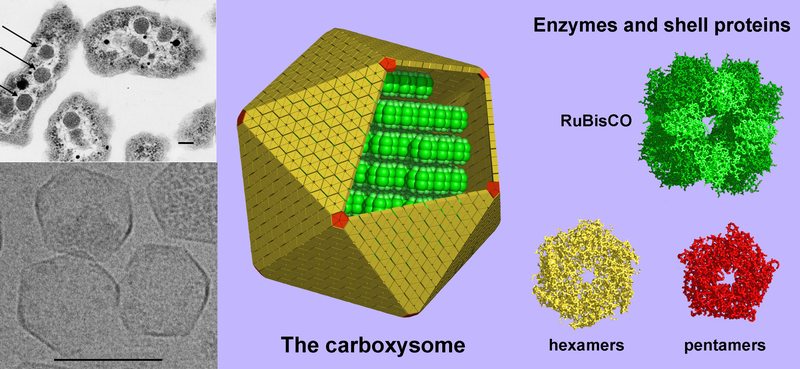
English: (Left, above) A thin-section electron micrograph of H. neapolitanus cells with carboxysomes inside. In one of the cells shown, arrows highlight the visible carboxysomes. (Left, below) Purified carboxysomes (material courtesy of S. Heinhorst and G. Cannon) as visualized by cryo-electron microscopy (courtesy of M. Yeager and K. Dryden). (right) Models for the structure of the carboxysome. Current data suggest that the shell is composed of several hundred hexameric protein building blocks and 12 pentameric building blocks. The three-dimensional atomic structures of the shell proteins have been determined by X-ray crystallography. RuBisCO, the main interior enzyme is shown packed inside in a regular arrangement for simplicity, though the actual organization of the enzymes is not understood yet. The other key enzyme, carbonic anhydrase, which is present in lesser amounts, is not illustrated. Scale bars are 100 nm. (image by T. Yeates). |
| Source | Modified version of Image:Carboxysome.png incorporating image taken from Image:Carboxysomes EM.jpg |
| Author | Prof. Todd O. Yeates, UCLA Dept. of Chem. and Biochem. |
| Other versions |
[]
PNG:
|


Licensing
[edit]
This file is licensed under the Creative Commons Attribution 3.0 Unported license.
- You are free:
- to share – to copy, distribute and transmit the work
- to remix – to adapt the work
- Under the following conditions:
- attribution – You must give appropriate credit, provide a link to the license, and indicate if changes were made. You may do so in any reasonable manner, but not in any way that suggests the licensor endorses you or your use.
File history
Click on a date/time to view the file as it appeared at that time.
| Date/Time | Thumbnail | Dimensions | User | Comment | |
|---|---|---|---|---|---|
| current | 01:38, 6 August 2008 | | 2,943 × 1,356 (3.72 MB) | TimVickers (talk | contribs) | {{Information |Description={{en|1=(Left, above) A thin-section electron micrograph of H. neapolitanus cells with carboxysomes inside. In one of the cells shown, arrows highlight the visible carboxysomes. (Left, below) Purified carboxysomes (material court |
You cannot overwrite this file.
File usage on Commons
The following 6 pages use this file:
File usage on other wikis
The following other wikis use this file:
- Usage on ar.wikipedia.org
- Usage on bg.wikipedia.org
- Usage on en.wikipedia.org
- Usage on en.wikiversity.org
- Usage on sw.wikipedia.org
- Usage on tr.wikipedia.org