File:Automated-Whole-Animal-Bio-Imaging-Assay-for-Human-Cancer-Dissemination-pone.0031281.s004.ogv
Jump to navigation
Jump to search
Size of this JPG preview of this OGG file: 600 × 600 pixels. Other resolutions: 240 × 240 pixels | 480 × 480 pixels | 768 × 768 pixels | 1,024 × 1,024 pixels.
Original file (Ogg Theora video file, length 1.4 s, 1,024 × 1,024 pixels, 1.17 Mbps, file size: 200 KB)
Captions
Captions
Add a one-line explanation of what this file represents
Summary
[edit]| Description |
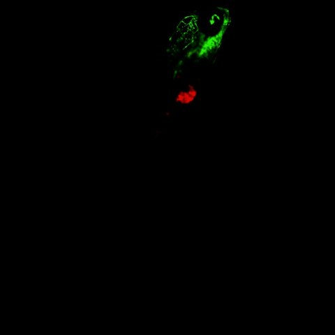
English: Combined multiple Z stacks of 6 dpi PC3-implanted embryo (range = 420 µm, step size = 30 µm, top = 180 µm, bottom = −210 µm). Red, CM-DiI-labeled tumor cells; green, GFP-endothelial cells of the Tg (Fli:GFP) line. |
||
| Date | |||
| Source | Video S1 from Ghotra V, He S, de Bont H, van der Ent W, Spaink H, van de Water B, Snaar-Jagalska B, Danen E (2012). "Automated Whole Animal Bio-Imaging Assay for Human Cancer Dissemination". PLOS ONE. DOI:10.1371/journal.pone.0031281. PMID 22347456. PMC: 3275564. | ||
| Author | Ghotra V, He S, de Bont H, van der Ent W, Spaink H, van de Water B, Snaar-Jagalska B, Danen E | ||
| Permission (Reusing this file) |
|
||
| Provenance |
|
File history
Click on a date/time to view the file as it appeared at that time.
| Date/Time | Thumbnail | Dimensions | User | Comment | |
|---|---|---|---|---|---|
| current | 09:30, 18 November 2012 | 1.4 s, 1,024 × 1,024 (200 KB) | Open Access Media Importer Bot (talk | contribs) | Automatically uploaded media file from Open Access source. Please report problems or suggestions here. |
You cannot overwrite this file.
File usage on Commons
There are no pages that use this file.