File:Amphioxus presumptive organ-forming areas in the uncleaved egg and durmg cleavage and blastulation.jpg
Original file (1,532 × 1,899 pixels, file size: 1.91 MB, MIME type: image/jpeg)
Captions
Captions
Summary
[edit]
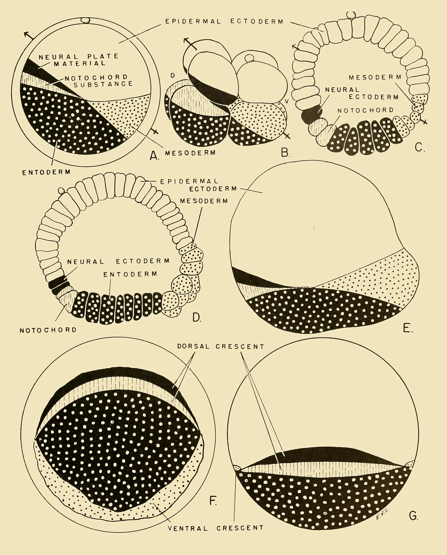
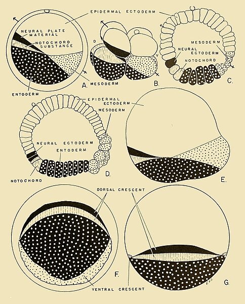
| Description | Fig 167 Presumptive organ-forming areas in the uncleaved egg and durmg cleavage and blastulation in Amphioxus. (Original diagram based upon data obtained from Conklin '32 '33.) (A) Uncleaved egg. (B) Eight-cell stage. (C) Early blastula in section (D) Late blastula in section. (E) Late blastula, external view from side. (F) Late blastula, external, vegetal pole view. (G) Late blastula, external, dorsoposterior view. The localization of cytoplasmic materials in Styela partita is similar to that of Amphioxus. Observe that the pointed end of the arrow defines the future cephalic end of the embryo. The position of the polar body denotes the antero-ventral area, while the position of the notochordal and neural plate material represents the antero-dorsal region. The "tail end" of the arrow is the postero-ventral area of the embryo. |
| Date | |
| Source | https://archive.org/details/comparativeembry00nels/page/335/mode/1up?view=theater&q=BLASTULATION+ Comparative embryology of the vertebrates; with 2057 drawings and photos. grouped as 380 illustrations. |
| Author | Nelsen, Olin E. |
Licensing
[edit]
|
This work is in the public domain in its country of origin and other countries and areas where the copyright term is the author's life plus 70 years or fewer.
| |
| This file has been identified as being free of known restrictions under copyright law, including all related and neighboring rights. | |

|
This file, which was originally posted to an external website, has not yet been reviewed by an administrator or reviewer to confirm that the above license is valid. See Category:License review needed for further instructions.
|
File history
Click on a date/time to view the file as it appeared at that time.
| Date/Time | Thumbnail | Dimensions | User | Comment | |
|---|---|---|---|---|---|
| current | 20:41, 11 March 2024 | | 1,532 × 1,899 (1.91 MB) | Rasbak (talk | contribs) | {{Information |description=Fig 167 Presumptive organ-forming areas in the uncleaved egg and durmg cleavage and blastulation in Amphioxus. (Original diagram based upon data obtained from Conklin '32 '33.) (A) Uncleaved egg. (B) Eight-cell stage. (C) Early blastula in section (D) Late blastula in section. (E) Late blastula, external view from side. (F) Late blastula, external, vegetal pole view. (G) Late blastula, external, dorsoposterior view. The localization of cytoplasmic materials in Styel... |
You cannot overwrite this file.
File usage on Commons
There are no pages that use this file.