Category:Blastula
Jump to navigation
Jump to search
sphere of cells formed during early embryonic development in animals | |||||
| Upload media | |||||
| Instance of |
| ||||
|---|---|---|---|---|---|
| Subclass of |
| ||||
| Follows | |||||
| Followed by |
| ||||
| |||||
Subcategories
This category has the following 2 subcategories, out of 2 total.
B
- Blastoderms (56 F)
- Blastodisc (5 F)
Media in category "Blastula"
The following 103 files are in this category, out of 103 total.
-
13227 2012 Article 55 Fig2 HTML.jpg 1,200 × 1,067; 121 KB
-
202211 Glass anemone blastula stage embryo.svg 1,000 × 1,000; 123 KB
-
-
Abatus cordatus Segmentation.jpg 1,640 × 1,592; 665 KB
-
American malacological bulletin (1986) (18156225785).jpg 2,358 × 2,106; 1.63 MB
-
Amia calva cleavage in the egg.jpg 1,002 × 827; 776 KB
-
Amphibia presumptive organ-forming areas in the late blastula and beginning gastrula.jpg 1,164 × 1,581; 1.61 MB
-
Amphioxus ovum segmentation.jpg 968 × 634; 629 KB
-
Amphioxus Three stages in the gastrulation.jpg 1,259 × 421; 389 KB
-
Animal forms; a second book of zoology (1902) (18008488228).jpg 2,336 × 2,700; 1.02 MB
-
Anodonta piscinalis segmentation embryo.jpg 1,287 × 379; 435 KB
-
Antedon rosacea early stages.jpg 801 × 735; 425 KB
-
Anthropogenie; oder, Entwickelungs-geschichte des Menschen (1910) (18746531984).jpg 1,856 × 2,632; 1.42 MB
-
Argyrotheca blastula postercolors.png 300 × 322; 11 KB
-
Ascaris Dorsal view of the segmenting egg of the 102-cell stage.jpg 800 × 754; 175 KB
-
Blastula (PSF) en rotate 05-ar.jpg 325 × 537; 45 KB
-
Blastula (PSF) en rotate 05.jpg 325 × 537; 78 KB
-
Blastula (PSF) rotada sp.JPG 337 × 539; 78 KB
-
Blastula (PSF).jpg 599 × 312; 33 KB
-
Blastula de xenopus.jpg 401 × 173; 30 KB
-
Blastula human.png 274 × 274; 32 KB
-
Blastula NWABR.jpg 2,560 × 1,920; 1.57 MB
-
Blastula ourico.jpg 1,216 × 294; 122 KB
-
Blastula polo animal.png 148 × 178; 51 KB
-
Blastula.jpg 241 × 273; 18 KB
-
Blastula.png 707 × 371; 171 KB
-
Blastulation.png 799 × 344; 100 KB
-
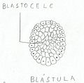
Blástula.jpg 608 × 599; 37 KB
-
Branchiostoma (YPM IZ 096163).jpeg 1,920 × 1,427; 532 KB
-
Cat longitudinal section through the axis of the ovum.jpg 878 × 706; 839 KB
-
Chordate Blastula fate map.PNG 290 × 290; 30 KB
-
Cinctoblastula type of development Homoscleromorpha.svg 977 × 673; 612 KB
-
Clytia Four stages in the development of the planula.jpg 1,306 × 582; 184 KB
-
Coeloblastula type of development Demospongiae.svg 978 × 305; 1.05 MB
-
Deuterostomes.png 1,992 × 927; 508 KB
-
Deuterostomia.png 590 × 263; 104 KB
-
Dierlijke ontwikkeling.png 2,798 × 966; 1.65 MB
-
Differentiation of the germ-track in Cyclops fuscus (A-H), and Eudiaptomus vulgaris (I).jpg 2,158 × 2,196; 1.05 MB
-
Dorsal lip transplantation in a salamander embryo.png 728 × 838; 649 KB
-
EB1911 Lamellibranchia - development of Ostrea edulis.jpg 775 × 985; 261 KB
-
EB1911 Sponges - Development of Sycon raphanus.jpg 925 × 1,092; 538 KB
-
EB1911 Tunicata - Stages in the Embryology of a Simple Ascidian.jpg 805 × 913; 216 KB
-
Ectopleura crocea. blastula (left) morula (right).jpg 1,074 × 825; 557 KB
-
Embryo in flower.png 3,000 × 3,006; 2.97 MB
-
Esquema de blastocele.jpg 594 × 441; 83 KB
-
Fertilization.jpg 1,920 × 1,080; 1.35 MB
-
Frog blastula blastocoel.jpg 2,208 × 1,092; 946 KB
-
Frog Segmentation of the egg and formation of the blastopore.jpg 1,111 × 1,212; 883 KB
-
Gastrulatie.png 1,780 × 1,520; 1.6 MB
-
Gastrulatsiooni toimumine.jpg 690 × 354; 67 KB
-
Gastrulação.png 695 × 349; 13 KB
-
-
-
-
Holothuria tubulosa development embryo.jpg 1,224 × 675; 776 KB
-
Human pedigree.jpg 2,329 × 2,723; 6.04 MB
-
Intercellular-Bridges-in-Vertebrate-Gastrulation-pone.0020230.s002.ogv 5.0 s, 417 × 202; 295 KB
-
Invaginacion1.png 333 × 214; 53 KB
-
Journal of morphology (1893) (14596154188).jpg 1,978 × 1,046; 177 KB
-
Journal of morphology (1893) Branchiostoma.jpg 1,071 × 1,617; 1.44 MB
-
La morphologie et stades developementaux de Nematostella.jpg 709 × 276; 51 KB
-
Lepidosiren paradoxa cleavage in the egg.jpg 925 × 648; 499 KB
-
Lumbricus Segmentation and early stages of development.jpg 1,124 × 1,088; 457 KB
-
Meyers b5 s0350 b1.png 570 × 218; 53 KB
-
Meyers b5 s0350.jpg 800 × 1,275; 412 KB
-
Meyers b5 s0682 b1.png 474 × 789; 304 KB
-
Midblastula embryo svg hariadhi.svg 512 × 512; 164 KB
-
Notopterus notopterus (10.3897-zse.93.13341) Figure 9.jpg 1,949 × 1,931; 1.95 MB
-
Ophiothrix fragilis early larvae 1.jpg 1,046 × 1,104; 424 KB
-
Ophiothrix fragilis early larvae 2.jpg 970 × 978; 413 KB
-
Ophiothrix fragilis early larvae.jpg 1,500 × 1,500; 583 KB
-
Paracentrotus lividus blastula.jpg 940 × 931; 738 KB
-
Paracentrotus lividus life cycle.jpg 725 × 693; 204 KB
-
Pedicellina echinata Early stages in the development of the egg.jpg 1,278 × 844; 239 KB
-
Petromyzon embryo (2).jpg 1,365 × 631; 203 KB
-
Platygaster hiemalis embryo development (00).jpg 671 × 1,151; 764 KB
-
Porifera- Generalized Amphiblastula Larva Settling.jpg 6,523 × 6,937; 4.23 MB
-
Protostomia.png 1,923 × 893; 582 KB
-
Protovsdeuterostomes-de.svg 577 × 527; 1.09 MB
-
Pseudorasbora parva (10.3897-zoologia.35.e22162) Figures 2–39.jpg 1,997 × 1,494; 1.42 MB
-
PSM V84 D534 Facts and factors of development fig10.jpg 595 × 767; 89 KB
-
Reptile blastula.jpg 744 × 606; 211 KB
-
Saccoglossus kowalevskii Blastula Gastrula.jpg 600 × 224; 66 KB
-
Schematic drawing of Calciblastula type of development Calcarea Calcinea.svg 1,056 × 697; 373 KB
-
Sea Urchin Blastula.jpg 762 × 676; 55 KB
-
Sea Urchin Vegetal Plate.jpg 698 × 546; 39 KB
-
Text-book of comparative anatomy (1898) (14777249844).jpg 1,204 × 1,404; 230 KB
-
The Biological bulletin (19757898313).jpg 1,998 × 2,332; 1.19 MB
-
The Biological bulletin (19758011533).jpg 2,004 × 1,310; 608 KB
-
The Biological bulletin (20187034358).jpg 1,700 × 2,098; 652 KB
-
The Biological bulletin (20190042950).jpg 1,908 × 2,880; 1.43 MB
-
The Biological bulletin (20190374640).jpg 2,004 × 2,406; 1.55 MB
-
The evolution of man (1920) plate III.jpg 753 × 1,173; 1.21 MB
-
Triturus viridescens cleavage.jpg 1,900 × 914; 509 KB
-
Vegetal rotation.png 1,000 × 1,680; 480 KB
-
Vegetal rotation.svg 512 × 860; 68 KB
-
Zfish midblastula stage embryo.jpg 1,118 × 1,102; 91 KB


