File:A-hybrid-blob-slice-model-for-accurate-and-efficient-detection-of-fluorescence-labeled-nuclei-in-3D-1471-2105-11-580-S11.ogv
Jump to navigation
Jump to search
Size of this JPG preview of this OGG file: 400 × 600 pixels. Other resolutions: 160 × 240 pixels | 512 × 768 pixels.
Original file (Ogg Theora video file, length 20 s, 512 × 768 pixels, 2.03 Mbps, file size: 4.87 MB)
Captions
Captions
Add a one-line explanation of what this file represents
Summary
[edit]| Description |
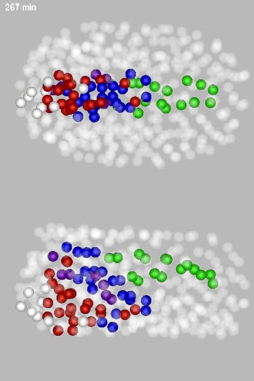
English: Movie 4: Precursors of the corpus and posterior bulb. 3D animation showing side by side left and ventral views of the embryo during the inflation stage of pharyngeal development. See legend of Figure 5II for coloring scheme. The movie illustrates the detailed reshaping of the pharynx from 197 through 337 minutes. In the later half of the assembly stage (197 to 250 minute), the two sheets expand in size through division. During the final round of synchronized divisions between ~277 and 307 the pharynx contracts along the AP axis rounding slightly. On the completion of divisions this structure then inflates to form a roundish structure[33] prior to its eventual elongating and spitting into two chambers (not shown). This ballooning is apparent from time 317 onward and occurs at the same time as the ventral MS pharynx cells move anteriorly toward the main mass of the pharynx. The movie also highlights the mouth precursors being born relatively distant from their final positions and converging near their final location. |
||
| Date | |||
| Source | Santella A, Du Z, Nowotschin S, Hadjantonakis A, Bao Z (2010). "A hybrid blob-slice model for accurate and efficient detection of fluorescence labeled nuclei in 3D". BMC Bioinformatics. DOI:10.1186/1471-2105-11-580. PMID 21114815. PMC: 3008706. | ||
| Author | Santella A, Du Z, Nowotschin S, Hadjantonakis A, Bao Z | ||
| Permission (Reusing this file) |
This file is licensed under the Creative Commons Attribution 2.0 Generic license.
|
||
| Provenance |
|
File history
Click on a date/time to view the file as it appeared at that time.
| Date/Time | Thumbnail | Dimensions | User | Comment | |
|---|---|---|---|---|---|
| current | 23:16, 4 June 2013 | 20 s, 512 × 768 (4.87 MB) | Open Access Media Importer Bot (talk | contribs) | Automatically uploaded media file from Open Access source. Please report problems or suggestions here. |
You cannot overwrite this file.
File usage on Commons
There are no pages that use this file.