File:59. Површинско набљудување на стомини клетки на листови од перуника (Iris germanica).ogg
Original file (Ogg multiplexed audio/video file, Theora/Vorbis, length 2 min 50 s, 1,920 × 1,080 pixels, 6.46 Mbps overall, file size: 131.01 MB)
Captions
Captions
Summary
[edit]| Description |
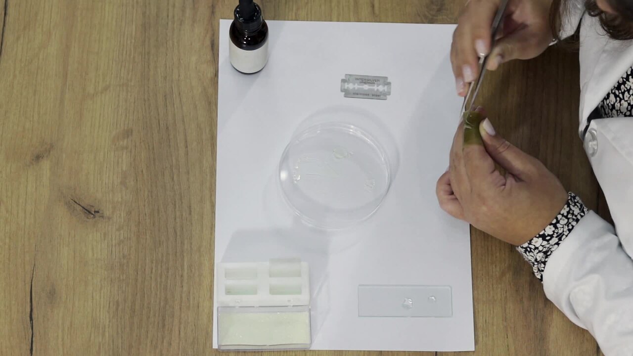
English: Surface observation of stomatal cells of leaves of bearded iris (Iris germanica) under microscope
Two drops of water are placed on the object glass, one central larger and one peripheral smaller. The iris leaf is taken. The leaf is placed around the index finger of the left hand. A thin cut is made with a razor blade, and then with tweezers and a razor blade, the epidermis is separated from the leaf. With the help of tweezers, the isolated piece of epidermis is placed in the central drop of water on the object glass. Then, a cover glass is placed on it, which is lowered next to the peripheral drop of water and at an angle of 45° it moves towards the middle of the object glass until the moment when the two drops of water merge, during which it is lowered. This procedure needs to be performed precisely because otherwise air bubbles will form which will count during microscopy. In this way the native microscopic slide is prepared and it is ready for microscopy. The epidermis is made up of basal epidermal cells, stomata cells and fibers. In the field of view of the microscope, the basic epidermal cells and the stomata are visible. The stomata in irises are built from two kidney-shaped stomata cells. Prepared and performed by Sara Cvetanoska, Institute of Biology, FNSM, Ss. Cyril and Methodius University, Skopje, Macedonia.Македонски: Површинско набљудување на стомини клетки на листови од перуника (Iris germanica) под микроскоп
Врз предметното стакленце се ставаат две капки вода, една средишна поголема и една странична помала капка вода. Се зема листот од перуника. Листот се става околу показалецот на левата рака. Се прави тенок пресек со помош на жилет, а потоа со пинцета и жилет се одвојува епидермисот од листот кој е во облик на тенка и проѕирна ципа. Со помош на пинцета изолираното парче од епидермис се става во средишната капка со вода на предметното стакленце. Потоа, се става покровно стакленце кое се спушта покрај страничната капка со вода и под агол од 45° се движи кон средината на предметното стакленце се до моментот кога ќе се спојат двете капки со вода, при што се спушта. Оваа постапка потребно е да се изведе прецизно затоа што во спротивно ќе се создадат воздушни меурчина кои ќе сметаат при микроскопирањето. На овој начин е подготвен нативниот микроскопски препарат и истиот е спремен за микроскопирање. Епидермисот е изграден од основни епидермални клетки, стомини клетки и влакна. Во видното поле на микроскоп се гледаат основните епидермални клетки и стомите. Стомите кај перуниката се изградени од две стомини клетки со бубреговиден облик. Подготвено и изведено од Сара Цветаноска, Институт за биологија, ПМФ, УКИМ, Скопје. |
| Date | |
| Source | Own work |
| Author | Deni Ingilizovski |

|
This biology experiment video was made within the Wikiexperiments 2022 set up by Shared Knowledge.
You can see all the videos in the category Biology Wikiexperiments.
|
Licensing
[edit]- You are free:
- to share – to copy, distribute and transmit the work
- to remix – to adapt the work
- Under the following conditions:
- attribution – You must give appropriate credit, provide a link to the license, and indicate if changes were made. You may do so in any reasonable manner, but not in any way that suggests the licensor endorses you or your use.
- share alike – If you remix, transform, or build upon the material, you must distribute your contributions under the same or compatible license as the original.
File history
Click on a date/time to view the file as it appeared at that time.
| Date/Time | Thumbnail | Dimensions | User | Comment | |
|---|---|---|---|---|---|
| current | 19:31, 15 November 2022 | 2 min 50 s, 1,920 × 1,080 (131.01 MB) | Dandarmkd (talk | contribs) | Uploaded own work with UploadWizard |
You cannot overwrite this file.
File usage on Commons
There are no pages that use this file.
Transcode status
Update transcode statusFile usage on other wikis
The following other wikis use this file:
- Usage on mk.wikipedia.org