Category:X-rays (radiation)
Jump to navigation
Jump to search
electromagnetic radiation of wavelength ranging from 10 pm to 10 nm | |||||
| Upload media | |||||
| Instance of | |||||
|---|---|---|---|---|---|
| Subclass of |
| ||||
| Part of | |||||
| Named after | |||||
| Discoverer or inventor | |||||
| Wavelength |
| ||||
| Follows | |||||
| Followed by | |||||
| Different from | |||||
| |||||
English: This category regards X-rays as an electromagnetic radiation. For pictures about Medical imaging, see Category Radiology or its subcategory Radiography.
Subcategories
This category has the following 23 subcategories, out of 23 total.
Pages in category "X-rays (radiation)"
The following 2 pages are in this category, out of 2 total.
Media in category "X-rays (radiation)"
The following 99 files are in this category, out of 99 total.
-
1896 caricature - The New Photographic Discovery.jpg 2,518 × 1,666; 1.07 MB
-
1plombs chasse cygne condé2.jpg 452 × 545; 105 KB
-
A schematic representation of mathematical relations in Fluctuation X-ray Scattering.jpg 2,998 × 2,248; 391 KB
-
Ampoule Puluj-12.jpg 87 × 128; 11 KB
-
Archives of physical medicine and rehabilitation (1920) (14756960595).jpg 1,326 × 424; 73 KB
-
Camadas electroes.png 497 × 445; 12 KB
-
Caméra XPAD3.png 260 × 359; 214 KB
-
Chocolat Guérin-Boutron. Dr Roentgen. Les bienfaiteurs de l'humanité.jpg 899 × 526; 464 KB
-
Clarendon Laboratory.jpg 2,756 × 2,397; 2.29 MB
-
CompoundRL-ru.svg 2,363 × 758; 2 KB
-
CompoundRL.jpg 881 × 280; 19 KB
-
Defense.gov photo essay 061020-D-7203T-008.jpg 4,056 × 2,640; 2.09 MB
-
Dereo UP kit.png 600 × 600; 194 KB
-
DeReO WA-P.png 200 × 200; 37 KB
-
Detecteur lineaire ligne a retard ambiguite photons symetriques.svg 335 × 140; 26 KB
-
Detecteur lineaire ligne a retard.svg 148 × 138; 25 KB
-
Discovery of X-rays by W.C. Röntgen.png 2,187 × 1,681; 33 KB
-
Dominant Photon-Matter Interaction.svg 608 × 401; 134 KB
-
Emise fluorescenčního záření.jpg 230 × 219; 11 KB
-
Emissão fóton raios x.png 678 × 316; 15 KB
-
Exposition Hautes Tensions - Tube a rayons X.JPG 2,592 × 1,944; 1.18 MB
-
ExpoSYFY - Terminator Salvation (10804025474).jpg 4,252 × 2,676; 7.81 MB
-
Farm-Fresh x ray.png 32 × 32; 3 KB
-
Fig 2 X-ray induced conductivity of ZnSe sensors.jpg 2,381 × 1,709; 358 KB
-
Fig 3 X-ray induced conductivity of ZnSe sensors.jpg 2,929 × 2,200; 568 KB
-
Fig 4 X-ray induced conductivity of ZnSe sensors.jpg 2,852 × 2,172; 375 KB
-
Filtratsiooni paksus - doos suhe.png 612 × 406; 39 KB
-
Font Awesome 5 solid x-ray.svg 512 × 410; 1 KB
-
Illuminated tram-car "Glasgow's X-ray campaign...", 1957 Wellcome L0016012.jpg 1,626 × 1,141; 924 KB
-
Ionisation chamber unscrewed to show components. Wellcome M0011488.jpg 4,126 × 2,808; 945 KB
-
JeanPerrin-Thèse-1897-Fig7.png 1,496 × 944; 183 KB
-
JeanPerrin-Thèse-1897-Fig9.png 1,088 × 585; 77 KB
-
Journal.pone.0149377.g001.PNG 2,862 × 3,271; 3.64 MB
-
Midland Tyre (as photographed by the Röntgen Rays).jpg 600 × 600; 100 KB
-
MiniFlex+ 1995.jpg 964 × 562; 216 KB
-
Multi-slice Acquisition Using Adaptive Array.jpg 640 × 480; 28 KB
-
NarrowBeamGeometry.jpg 731 × 396; 50 KB
-
Optical layout RIXS spectrometer.png 560 × 683; 44 KB
-
OSI Layers.png 560 × 332; 46 KB
-
Painted Lady Chrysalis micro CT.jpg 4,175 × 2,674; 2.5 MB
-
Parasite170054-fig5 Rhadinorhynchus oligospinosus (Acanthocephala).png 2,126 × 2,433; 1.59 MB
-
Parasite170054-fig6 Rhadinorhynchus oligospinosus (Acanthocephala).png 2,126 × 2,420; 1.66 MB
-
Pictor A xray.jpg 864 × 864; 132 KB
-
Portrait of Sir James Mackenzie Davidson Wellcome M0013472.jpg 2,785 × 3,932; 3.15 MB
-
Principles of X-ray generation.jpg 800 × 620; 57 KB
-
Propagation-based imaging.PNG 399 × 249; 9 KB
-
Pump probe x ray spectroscopy.png 888 × 461; 269 KB
-
RadioactiveImage.png 1,920 × 1,080; 2 MB
-
RadWiki-Condense-Facts-and-Information-to-Knowledge.png 1,200 × 848; 41 KB
-
Rbe definition.png 491 × 410; 18 KB
-
Refletometria de Raios X em filmes finos.jpg 501 × 393; 35 KB
-
Rixs cartoon.png 3,232 × 1,819; 3.39 MB
-
Roentgenbeugung in einer Fluessigkeit.svg 381 × 276; 80 KB
-
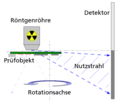
Rotations-Laminographie 1.png 907 × 535; 77 KB
-
Rowland circle geometry.png 1,065 × 717; 118 KB
-
Rowland Circle with two orders.png 1,028 × 848; 90 KB
-
Röntgenstrahlung (Erklärvideo des Bundesamt für Strahlenschutz).webm 5 min 2 s, 640 × 360; 6.98 MB
-
Röntgenstrålning - Ystad-2021.jpg 1,742 × 1,111; 1.84 MB
-
San Domenico31.jpg 982 × 902; 428 KB
-
Schema HRCL.png 489 × 423; 43 KB
-
Schematic IXS spectrum.png 1,187 × 783; 669 KB
-
Sediment-core PS56 039 x-ray hg.jpg 1,472 × 3,000; 956 KB
-
Sediment-images hg.jpg 734 × 525; 205 KB
-
Size Changed 20171016 110937170 CXI.jpg 720 × 1,080; 464 KB
-
StrukturierteSzintillatoren.jpg 564 × 288; 25 KB
-
SWI negative phase mask.png 908 × 380; 30 KB
-
Systematik Strahlenschäden.PNG 959 × 719; 25 KB
-
TBKPhoto.jpg 1,048 × 1,360; 506 KB
-
TFTArrayauslesen.jpg 907 × 596; 48 KB
-
The complete disintegration series of uranium. Wellcome M0015376.jpg 2,808 × 3,954; 952 KB
-
Threshold LUT.gif 495 × 385; 22 KB
-
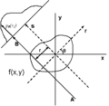
Tomographic fig1.png 400 × 390; 5 KB
-
Tomographic Imaging Labels.jpg 401 × 350; 57 KB
-
Tomographic imaging slice selection.jpg 512 × 294; 15 KB
-
Translatorial Laminography.png 858 × 661; 56 KB
-
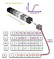
Volume Rendering Up.jpg 564 × 624; 41 KB
-
X-ray (1921) (14767179591).jpg 1,028 × 1,698; 419 KB
-
X-ray (1922) (14801933623).jpg 1,280 × 1,968; 652 KB
-
X-ray free-space propagation - from near field to far field.png 960 × 720; 138 KB
-
X-Ray region of the electromagnetic spectrum.jpg 2,494 × 350; 97 KB
-
X-ray vision, peeking into the atomic world.jpg 1,146 × 1,432; 503 KB
-
X-Riot.jpg 500 × 500; 39 KB
-
XPS CC Roland Siegbert.svg 1,052 × 744; 703 KB
-
Xray-phase-contrast-disks-color.png 500 × 500; 3 KB
-
Xray-phase-contrast-disks0000mm.jpg 500 × 500; 5 KB
-
Xray-phase-contrast-disks1000mm.jpg 500 × 500; 25 KB
-
XrayAF.jpg 660 × 503; 52 KB


